BIC (@bic_bordeaux) 's Twitter Profile
BIC

@bic_bordeaux

Bordeaux Imaging Center

ID: 1968536221

linkhttp://www.bic.u-bordeaux.fr/ calendar_today18-10-2013 09:10:54

473 Tweet

1,1K Followers

378 Following

BIC (@bic_bordeaux) 's Twitter Profile Photo

Second FBI talk this morning as part of the #RIME meeting: latests news about the development of a MOOC dedicated to ultramicrotomy as part of #FBITraining

Second FBI talk this morning as part of the #RIME meeting: latests news about the development of a MOOC dedicated to ultramicrotomy as part of #FBITraining
BIC (@bic_bordeaux) 's Twitter Profile Photo

#ImageOftheMonth Hélène Bonnet INRAE UMR1332 BFP In June say it with flowers. This image, taken under a macroscope at the plant unit of the BIC, shows flower bud from the sweet cherry tree ‘Burlat’, sampled in the orchard and grown at 24°C for ten days to observe flowering.

#ImageOftheMonth
Hélène Bonnet <a href="/INRAE_BFP/">INRAE UMR1332 BFP</a> 
In June say it with flowers.
This image, taken under a macroscope at the plant unit of the BIC, shows flower bud from the sweet cherry tree ‘Burlat’, sampled in the orchard and grown at 24°C for ten days to observe flowering.
BIC (@bic_bordeaux) 's Twitter Profile Photo

After 6 month of grey sky, today we try to convince the sun to be present and to stay with us! Here comes the sun, here is the Yellow BIC !

After 6 month of grey sky, today we try to convince the sun to be present and to stay with us! Here comes the sun, here is the Yellow BIC !
BIC (@bic_bordeaux) 's Twitter Profile Photo

Today @JTeillon and Poujol Christel are opening the workshop sessions of the photonic unit with a full day talking about Tissue optical clearing.

Today @JTeillon and <a href="/ChristelPoujol/">Poujol Christel</a> are opening the workshop sessions of the photonic unit with a full day talking about Tissue optical clearing.
BIC (@bic_bordeaux) 's Twitter Profile Photo

#ImageOfTheMonth July image from Angela Getz Interdisciplinary Institute for Neuroscience 🧠 shows a sparse labeling of endogenous AMPA receptors on CA1 pyramidal neuron dendrites in the stratum radiatum. 3D image was acquired using lattice light-sheet microscopy.

#ImageOfTheMonth
July image from <a href="/ang_getz/">Angela Getz</a> <a href="/iins_bordeaux/">Interdisciplinary Institute for Neuroscience 🧠</a> shows a sparse labeling of endogenous AMPA receptors on CA1 pyramidal neuron dendrites in the stratum radiatum. 3D image was acquired using lattice light-sheet microscopy.
France-BioImaging Research Infrastructure (@fr_bioimaging) 's Twitter Profile Photo

Curious about the benefits of the cryoCLEM technology & how to apply it to your research project? This workshop covers the entire workflow from sample preparation to correlative microscopy in cryogenic conditions🔬 📍BIC 📆October 14-15, 2024 france-bioimaging.org/announcement/c…

Curious about the benefits of the cryoCLEM technology &amp; how to apply it to your research project? This workshop covers the entire workflow from sample preparation to correlative microscopy in cryogenic conditions🔬 
📍<a href="/BIC_Bordeaux/">BIC</a> 
📆October 14-15, 2024
france-bioimaging.org/announcement/c…
Luis AJ Alvarez (@l_aj_alvarez) 's Twitter Profile Photo

Poujol Christel & Magali Grison from BIC running a hands on session in TauSTED in plants with both multicolour fixed but also live plants looking at Golgi organisation #EMBLSuperResolution

<a href="/ChristelPoujol/">Poujol Christel</a> &amp; Magali Grison from <a href="/BIC_Bordeaux/">BIC</a> running a hands on session in TauSTED in plants with both multicolour fixed but also live plants looking at Golgi organisation  #EMBLSuperResolution
BIC (@bic_bordeaux) 's Twitter Profile Photo

#ImageOfTheMonth August image from M.Brando ISVV - Institut des Sciences de la Vigne et du Vin & I.Svahn BIC shows pollen and mildew on the stamen stem of a vine flower. It was obtained using Cryo Scanning Electron Microscopy.

#ImageOfTheMonth
August image from M.Brando <a href="/isvv_bordeaux/">ISVV - Institut des Sciences de la Vigne et du Vin</a> &amp; I.Svahn <a href="/BIC_Bordeaux/">BIC</a> shows pollen and mildew on the stamen stem of a vine flower. It was obtained using Cryo Scanning Electron Microscopy.
BIC (@bic_bordeaux) 's Twitter Profile Photo

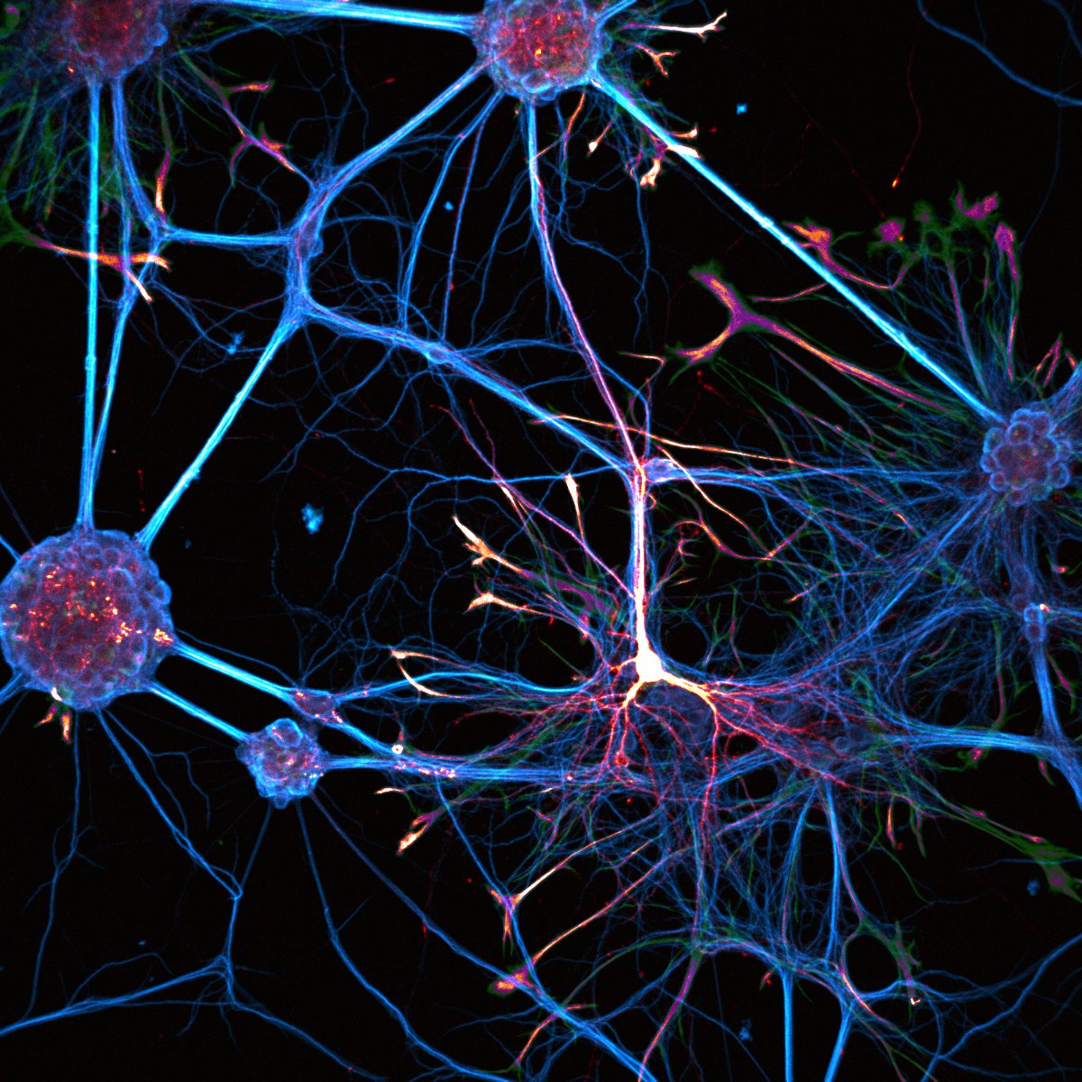
#ImageOfTheMonth This image was acquired by J.Angibaud Interdisciplinary Institute for Neuroscience 🧠. Neurons and astrocytes are cultured on a soft polyacrylamide hydrogel. Neurons are labeled for MAP-2 protein (blue), astrocytes for GFAP protein (red) and transfected neurons show GFP expression (green).
#ImageOfTheMonth
This image was acquired by J.Angibaud <a href="/iins_bordeaux/">Interdisciplinary Institute for Neuroscience 🧠</a>. Neurons and astrocytes are cultured on a soft polyacrylamide hydrogel. Neurons are labeled for MAP-2 protein (blue), astrocytes for GFAP protein (red) and transfected neurons show GFP expression (green).
Daniel Choquet (@daniel_choquet1) 's Twitter Profile Photo

Bordeaux Imaging Center, la fabrique bordelaise d’images scientifiques célèbre ses 15 ans | Délégation Aquitaine aquitaine.cnrs.fr/fr/cnrsinfo/bo…

BIC (@bic_bordeaux) 's Twitter Profile Photo

This year, BIC is organizing the annual meeting of INRAE microscopist network with seminars and workshops (mostly in french). Registration are still open: rmui2024.journees.inrae.fr

This year, BIC is organizing the annual meeting of <a href="/INRAE_France/">INRAE</a> microscopist network with seminars and workshops (mostly in french). Registration are still open: rmui2024.journees.inrae.fr
BIC (@bic_bordeaux) 's Twitter Profile Photo

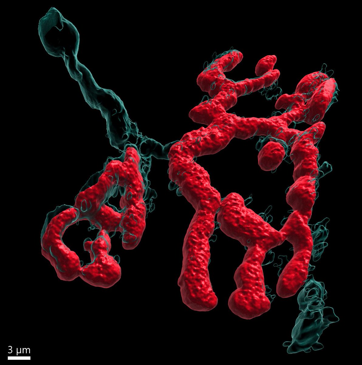
#ImageOfTheMonth TOURNEZY Jeflie from Neurocentre Magendie, helped by M. Fernández Monreal BIC made this 3D reconstruction Imaris 3D/4D Imaging of a mouse extensor digitorum longus neuromuscular junction. In cyan we can see the pre-synaptic terminal and in red the post-synaptic motor plate.
#ImageOfTheMonth
<a href="/TJeflie/">TOURNEZY Jeflie</a> from <a href="/NCMagendie/">Neurocentre Magendie</a>, helped by M. Fernández Monreal <a href="/BIC_Bordeaux/">BIC</a> made this 3D reconstruction <a href="/ImarisSoftware/">Imaris 3D/4D Imaging</a> of a mouse extensor digitorum longus neuromuscular junction. In cyan we can see the pre-synaptic terminal and in red the post-synaptic motor plate.
France-BioImaging Research Infrastructure (@fr_bioimaging) 's Twitter Profile Photo

We are at #GBI_EoE2024! 🇯🇵 Very glad to be part of this 9th edition focusing on democratizing access to image data management tools & resources 🔬 Looking forward to discussing with the imaging community around the world! 🌍 Global BioImaging

We are at #GBI_EoE2024! 🇯🇵 
Very glad to be part of this 9th edition focusing on democratizing access to image data management tools &amp; resources 🔬
Looking forward to discussing with the imaging community around the world! 🌍
<a href="/GlobalBioImage/">Global BioImaging</a>
France-BioImaging Research Infrastructure (@fr_bioimaging) 's Twitter Profile Photo

[Event] It's a wrap for the FBI Annual Meeting 2024! For the last day, live functional #imaging was in the spotlight with 2 keynotes from major players in this field and many presentations. A big thank you to everyone who joined and contributed to this inspiring event🙌

[Event] It's a wrap for the FBI Annual Meeting 2024!

For the last day, live functional #imaging was in the spotlight with 2 keynotes from major players in this field and many presentations.

A big thank you to everyone who joined and contributed to this inspiring event🙌