Cambridge Advanced Imaging Centre
@cammicroscopy
Advanced imaging research facility in the School of Biological Sciences at the University of Cambridge caic.bio.cam.ac.uk
ID: 1063449674961625095
16-11-2018 15:12:15
30 Tweet
450 Followers
114 Following


Flaminia Kaluthantrige Don, a Biotechnology and Biological Sciences Research CASE student hosted by CAIC and Meri Huch's lab, used our two-photon microscope to great effect. She optimised imaging conditions in order to observe single cells proliferating into liver organoids over three days.

Great work by Martin Lenz showcased in his talk about the multi-view light sheet microscope for gentle fast imaging of plants he has developed in the Sainsbury Laboratory Cambridge University (SLCU) imaging facility.


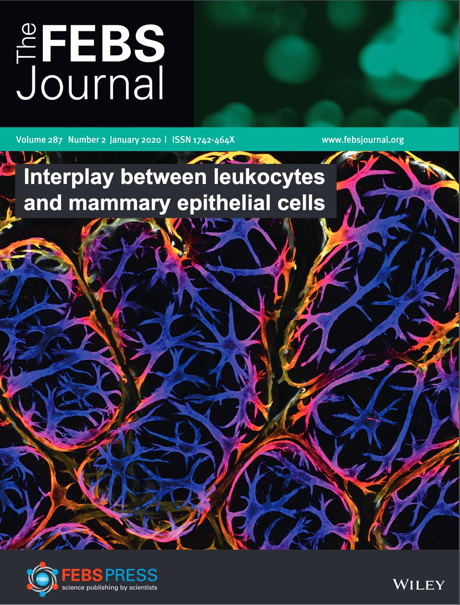
We got the cover! The wonderful Jessie Hitchcock's new paper is out now in The FEBS Journal, with an excellent commentary from Dr Wendy Ingman. If you like immune cells and boobs, or nice 3D imaging, this is worth a read. febs.onlinelibrary.wiley.com/doi/10.1111/fe… with Kate Hughes & Christine J Watson


The Sarris Lab made incredible use of our two-photon microscope in their recently published research on neutrophil migration to sites of tissue damage cell.com/current-biolog…


August image of the month: Drosophila larvae expressing green and magenta fluorescent proteins in nociceptive (noxious touch), and proprioceptive (body movement) neurons. Image: Paul Brooks who researches neuron degeneration, captured by confocal microscope in Dept of Zoology


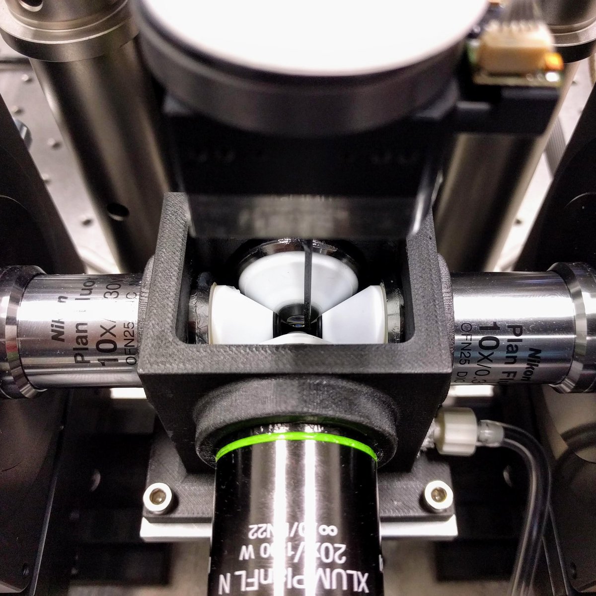
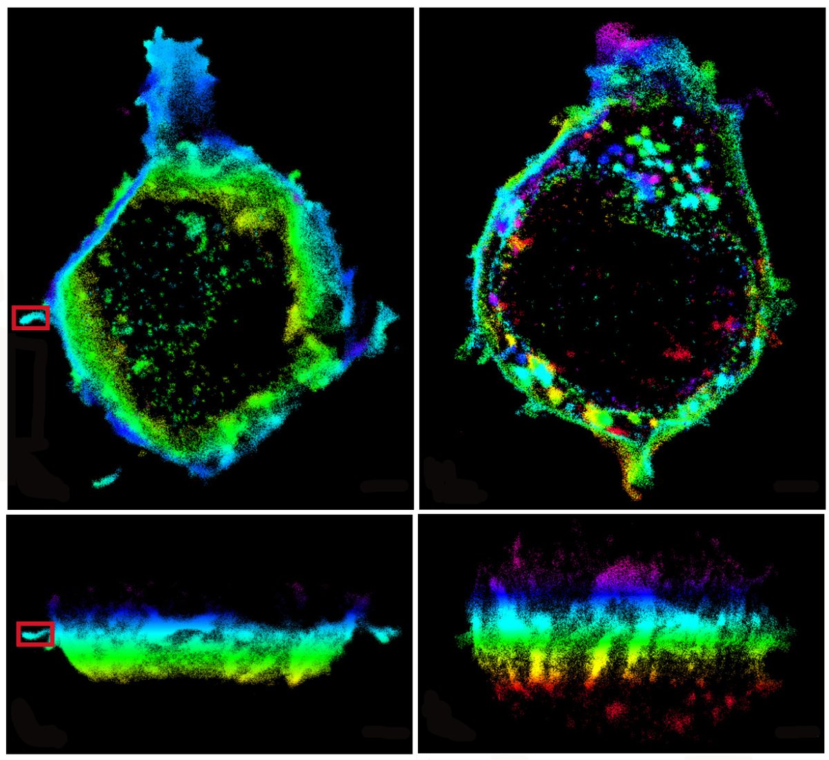
Via #OSA_Optica: Single molecule light field microscopy ow.ly/Urg550BEkQ4 #FlatOptics #SpectralFIlters @koholleran Cambridge University University of Leeds


how to cover yourself when you are a plant? Just weave your own fabric! Check our collaborative work between Sainsbury Laboratory Cambridge University (SLCU) Cam Botanic Garden Cambridge Chemistry Cambridge Advanced Imaging Centre on how Dionysia tapetodes extrudes flavones threads from its glandular trichomes. biorxiv.org/content/10.110…

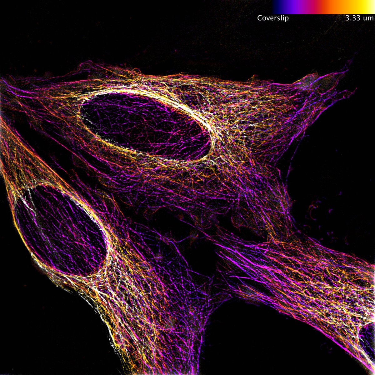
October image of the month is from Dr. Ruby Peters of the Ewa Paluch 🇺🇦 and shows HeLa cells, labelled with Tubulin-GFP. Taken using our Elyra 7 Lattice SIM and colour coded for depth


Check out our latest publication 'Visualising an invisible symbiosis' in Plants People Planet 🌱👥🌐! Department of Plant Sciences Crop Science Centre Niab nph.onlinelibrary.wiley.com/doi/full/10.10…

A great opportunity in Cambridge to lead the Advanced Imaging Centre at the School of Biological Science ... 10 more days to apply jobs.cam.ac.uk/job/40627/ Cambridge University School of Biological Sciences RMS Euro-BioImaging ERIC FocalPlane


