Graef Lab (@graeflab) 's Twitter Profile
Graef Lab

@graeflab

We are interested in the #cell_biology of #autophagy, #mitochondria, and #ageing.

ID: 1212848842414022657

linkhttps://www.age.mpg.de/science/research-laboratories/graef/ calendar_today02-01-2020 21:31:23

516 Tweet

1,1K Followers

362 Following
mito911 (@mito911_) 's Twitter Profile Photo

Congrats to Dr. Shanlin Rao Hummer Lab et al. for revealing in high detail the gymnastic of the LC3 lipidation machinery as it prepares autophagic membranes for cargo engagement using MD simulations, in vitro reconstitution and cell biology science.org/doi/10.1126/sc…
Michael Lazarou (@lazaroulab) 's Twitter Profile Photo

Molecular simulations, biochemistry, and cell biology come together to reveal how a crucial step in autophagy is executed. The work was made possible through a fantastic synergy between three labs, including ours, located in different continents across the world.

Sho Suzuki (he/him) (@shosuzuki20) 's Twitter Profile Photo

I am super excited to share that my first corresponding author paper has been published in JCB (Journal of Cell Biology). Surprising role of ER in ESCRT-mediated sorting! rupress.org/jcb/article/22… (1/8)

I am super excited to share that my first corresponding author paper has been published in JCB (<a href="/JCellBiol/">Journal of Cell Biology</a>). Surprising role of ER in ESCRT-mediated sorting! rupress.org/jcb/article/22… (1/8)
Patrick Lusk (@plusk4u) 's Twitter Profile Photo

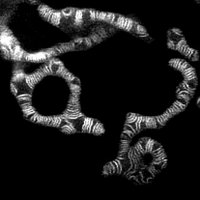
Dynamin-like proteins at the nuclear envelope? That and other surprises in excellent work by Philip Mannino with help from Nick Ader Melia Lab and @LuskingL! #autophagy biorxiv.org/content/10.110…

JakobsLab (@jakobslab) 's Twitter Profile Photo

The human prohibitin complex forms a bell-shaped structure in the inner mitochondrial membrane. Check out our #preprint together with Michael Ratz below 👇 biorxiv.org/content/10.110… What do you think it looks like? Like a bundt cake? A jellyfish? Vanilla pudding?😉

Maria Bohnert (@mariabohnert) 's Twitter Profile Photo

I am thrilled to see our work on the #Vacuole #LipidDroplet #MembraneContactSite vCLIP published in Developmental Cell! Fantastic collaboration with Ruben Fernandez and @schmidtlabIBK. Huge thanks to the whole team, I am so proud of you all! sciencedirect.com/science/articl…
AUTOPHAGY-AARHUS (Reggiori & Mari Research Labs) (@autophagyaarhus) 's Twitter Profile Photo

We are happy to share the publication below, which sheds light on the role of Atg1, Atg11 and Atg13 in bulk autophagy initiation. You can find the paper in the following link 👇 pubmed.ncbi.nlm.nih.gov/38233718/

We are happy to share the publication below, which sheds light on the role of Atg1, Atg11 and Atg13 in bulk autophagy initiation. You can find the paper in the following link 👇
pubmed.ncbi.nlm.nih.gov/38233718/
Weill Institute for Cell & Molecular Biology (@weillinstitute) 's Twitter Profile Photo

🚨The Weill Institute is hiring a new faculty member!🚨 Assistant Professor position is now open in Weill Institute for Cell & Molecular Biology, joint with Cornell MBG. This is an open search in all areas of molecular and cell biology; application deadline is 3/20. More info: academicjobsonline.org/ajo/jobs/27253

Wollert Lab (@wollertlab) 's Twitter Profile Photo

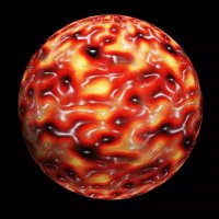
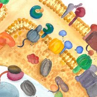
Eat less to live longer? #Autophagy degrades damaged or toxic cellular material to keep cells in good shape. We discovered how #ATG16L1 shapes phagophores. Our article was published today in NatureStructMolBiol ! thanks to all the authors !! Check it out: nature.com/articles/s4159…

Eat less to live longer? #Autophagy degrades damaged or toxic cellular material to keep cells in good shape. We discovered how #ATG16L1 shapes phagophores. Our article was published today in <a href="/NatureSMB/">NatureStructMolBiol</a> ! thanks to all the authors !! Check it out: 
nature.com/articles/s4159…
OsmanLab (@osman_lab) 's Twitter Profile Photo

🚀Excited to share our latest paper, where we explore dynamic changes in mtDNA heteroplasmy in living cells! Discover how asymmetric partitioning and mitochondrial dynamics drive mtDNA variant segregation! 🔬embopress.org/doi/full/10.10… #Mitochondria #microscopy #microfluidics #mtDNA

Rupert Beale (@bealelab) 's Twitter Profile Photo

We’ve worked out how the molecular machines that create acidic compartments can recruit autophagy (self-eating) machinery when they can’t maintain a pH gradient. We are really pleased with this discovery, out in Molecular Cell cell.com/molecular-cell…. 1/16
Cornell MBG (@cornellmbg) 's Twitter Profile Photo

Excited to announce an MBG faculty search for two assistant professor positions in collaboration with the Weill Institute for Cell and Molecular Biology, Weill Institute for Cell & Molecular Biology. Please share and apply! academicjobsonline.org/ajo/jobs/28336

Ragusa Lab (@ragusalab) 's Twitter Profile Photo

Our work on the biochemical reconstitution of mitophagy initiation led by devikaA has been updated on bioRxiv. You can check it out at biorxiv.org/content/10.110…

Our work on the biochemical reconstitution of mitophagy initiation led by <a href="/Devika_An/">devikaA</a>  has been updated on bioRxiv. You can check it out at biorxiv.org/content/10.110…
Kraft Lab (@kraftlabfr) 's Twitter Profile Photo

🎉 We're thrilled to share that our former colleague, Mariya Licheva, now with the Graef Lab, received the prestigious Hans-Grisebach-Preis from Universität Freiburg for her dissertation! 🏅Congrats on this great achievement, Mariya! Photo credit: Universität Freiburg / Jürgen Gocke

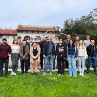
🎉 We're thrilled to share that our former colleague, <a href="/_mariyalicheva/">Mariya Licheva</a>, now with the <a href="/GraefLab/">Graef Lab</a>, received the prestigious Hans-Grisebach-Preis from <a href="/UniFreiburg/">Universität Freiburg</a> for her dissertation! 🏅Congrats on this great achievement, Mariya!

Photo credit: Universität Freiburg / Jürgen Gocke
Hurley Lab (@thehurleylab) 's Twitter Profile Photo

Proud of new work tinyurl.com/yhz4c6ry from the lab by Zhicheng (Chen) Cui Zhicheng Cui | 崔志成 using single-particle cryo-EM on liposomes to show how mTORC1 integrates nutrient and growth factor signaling at lysosomes, collab. w/ Gennaro Napolitano, A. Esposito, and A. Ballabio.

Proud of new work tinyurl.com/yhz4c6ry from the lab by Zhicheng (Chen) Cui <a href="/Chen3dem/">Zhicheng Cui | 崔志成</a> using single-particle cryo-EM on liposomes to show how mTORC1 integrates nutrient and growth factor signaling at lysosomes, collab. w/ <a href="/G__Napolitano/">Gennaro Napolitano</a>, A. Esposito, and A. Ballabio.