John Brealey (@johnbrealey) 's Twitter Profile
John Brealey

@johnbrealey

Senior Medical Scientist, Electron Microscopy Unit, SA Pathology, Adelaide, Australia. Ultrastructural pathology of kidney, muscle, nerve, liver, tumours, etc.

ID: 1121386760397713409

calendar_today25-04-2019 12:13:32

881 Tweet

1,1K Followers

1,1K Following

John Brealey (@johnbrealey) 's Twitter Profile Photo

Kidney Bx. Patient has monoclonal immunoglobulin deposition disease (MIDD). #electronmicroscopy shows a macrophage in close apposition with a tubular basement membrane (TBM). The macrophage appears to be processing the powdery 'punctate' electron-dense deposit within the TBM.

Kidney Bx. Patient has monoclonal immunoglobulin deposition disease (MIDD). #electronmicroscopy shows a macrophage in close apposition with a tubular basement membrane (TBM). The macrophage appears to be processing the powdery 'punctate' electron-dense deposit within the TBM.
John Brealey (@johnbrealey) 's Twitter Profile Photo

#askrenal Tiffany Caza Lynn D. Cornell, M.D. Child w heart tx. Native kidney bx. Proteinuria + hematuria. C3 + C4 (N), anti-GBM (+), dsDNA (-), cryo (-), Congo Red (-), DNAJB9 (-), IgG 1-2+ linear, C3 (weak). EM: focal subepithelial organized dep in a membranous pattern. Occ TRI. Any ideas?
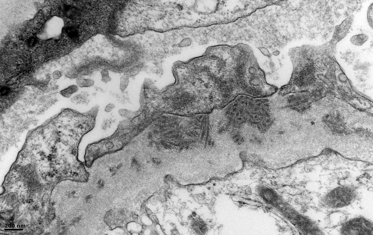
#askrenal <a href="/Tiff_Caza/">Tiffany Caza</a> <a href="/ldcornell/">Lynn D. Cornell, M.D.</a> Child w heart tx. Native kidney bx. Proteinuria + hematuria. C3 + C4 (N), anti-GBM (+), dsDNA (-), cryo (-), Congo Red (-), DNAJB9 (-), IgG 1-2+ linear, C3 (weak). EM: focal subepithelial organized dep in a membranous pattern. Occ TRI. Any ideas?
John Brealey (@johnbrealey) 's Twitter Profile Photo

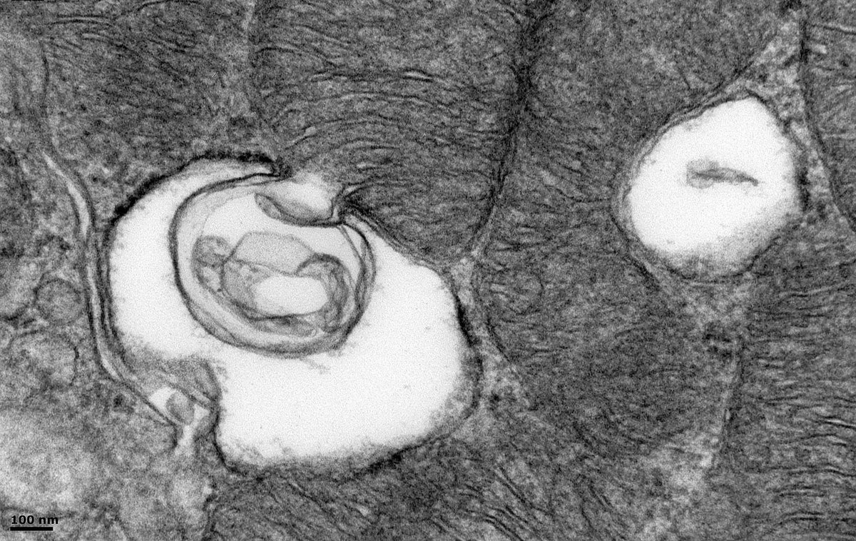
Kidney Bx #electronmicroscopy shows tubular epithelium. What's going on with the mitochondria? It looks like the mitochondrion is being internalised within a phagophore. You can see the cristae being 'sucked in' to the structure. I interpret this as mitophagy - is this correct?

Kidney Bx #electronmicroscopy shows tubular epithelium. What's going on with the mitochondria? It looks like the mitochondrion is being internalised within a phagophore. You can see the cristae being 'sucked in' to the structure. I interpret this as mitophagy - is this correct?
John Brealey (@johnbrealey) 's Twitter Profile Photo

#askrenalpath Astrid Weins Do patients with anti-nephrin autoantibodies still show slit diaphragms between intact podocyte foot processes by #electronmicroscopy?

John Brealey (@johnbrealey) 's Twitter Profile Photo

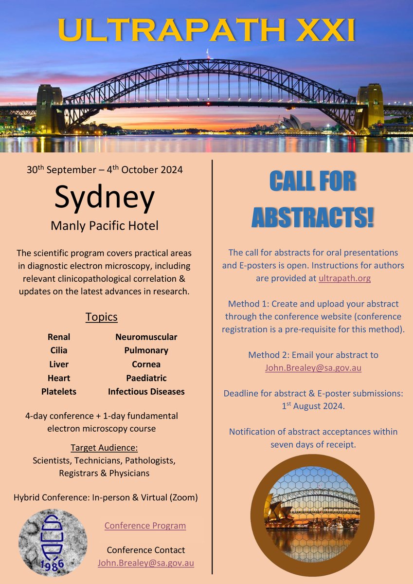
Please consider attending the UltraPath XXI electron microscopy conference in beautiful Manly Beach, Sydney, Sep 30 to Oct 4, 2024. Lots of interesting ultrastructural pathology with presentations by world leaders in the field. Plus great social events. ultrapath.org

Please consider attending the UltraPath XXI electron microscopy conference in beautiful Manly Beach, Sydney, Sep 30 to Oct 4, 2024. Lots of interesting ultrastructural pathology with presentations by world leaders in the field. Plus great social events.

ultrapath.org
John Brealey (@johnbrealey) 's Twitter Profile Photo

Seeking opinions on this subendothelial material. Native Renal Bx. Male in 60s w hematuria + proteinuria. LM shows an MPGN w focal vasculitis. EM is from the paraffin block - occasional loops occluded. IF - focal staining for fibrinogen. ? cryofibrinogen #askrenal Sanjeev Sethi
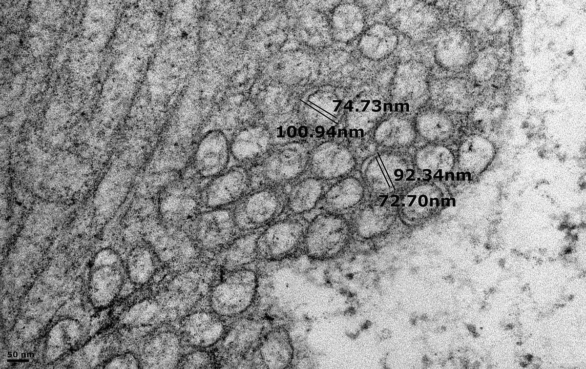
Seeking opinions on this subendothelial material. Native Renal Bx. Male in 60s w hematuria + proteinuria. LM shows an MPGN w focal vasculitis. EM is from the paraffin block - occasional loops occluded. IF - focal staining for fibrinogen. ? cryofibrinogen
#askrenal <a href="/SethiRenalPath/">Sanjeev Sethi</a>
John Brealey (@johnbrealey) 's Twitter Profile Photo

Seeking opinions for a differential on this appearance by #electronmicroscopy. Native Renal Bx, male in 30s. My thoughts are LCAT Deficiency or Alagille Syndrome. Any others? - ? hepatic glomerulosclerosis. Pt has low HDL. #askrenal Sanjeev Sethi Anthony Chang, MD (張賀文) Jonathan Zuckerman MD PhD
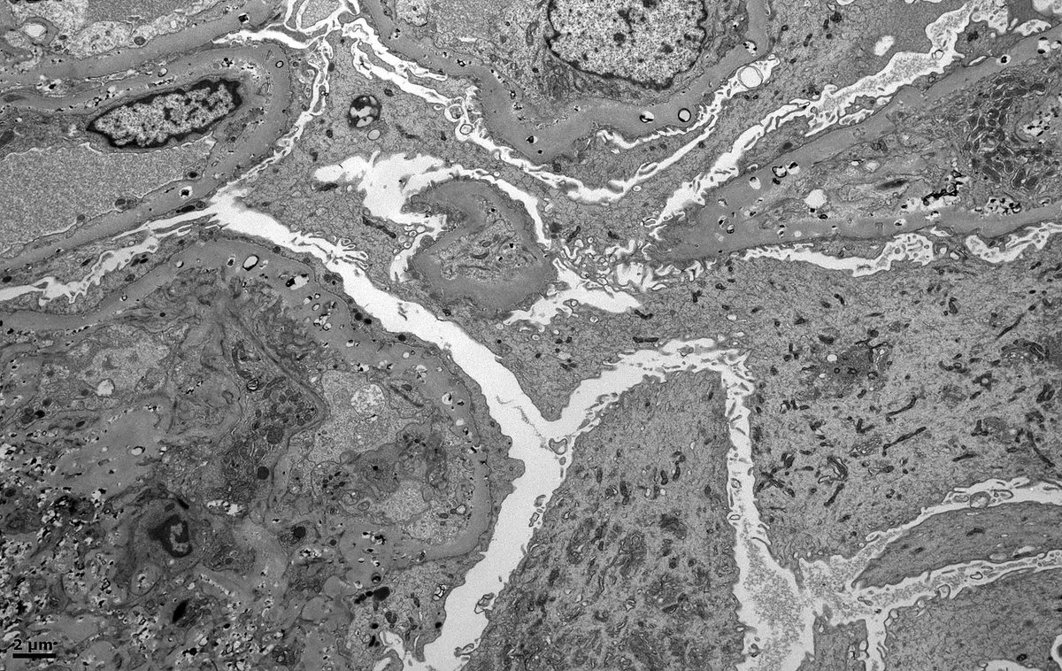
Seeking opinions for a differential on this appearance by #electronmicroscopy. Native Renal Bx, male in 30s. 
My thoughts are LCAT Deficiency or Alagille Syndrome. Any others? - ? hepatic glomerulosclerosis. Pt has low HDL. #askrenal <a href="/SethiRenalPath/">Sanjeev Sethi</a> <a href="/ChangUCanSpare/">Anthony Chang, MD (張賀文)</a> <a href="/JZRenalPath/">Jonathan Zuckerman MD PhD</a>
John Brealey (@johnbrealey) 's Twitter Profile Photo

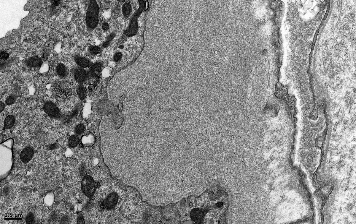
Renal Bx, female in 40s. TBMs focally expanded by fibrils measuring 12-13nm in diameter. I see this occasionally by EM in renal biopsies and never know if it's amyloid or a mimic (similar to diabetic fibrillosis in mesangial matrix). No amyloid in gloms. Congo Red not done ATM.

Renal Bx, female in 40s. TBMs focally expanded by fibrils measuring 12-13nm in diameter. I see this occasionally by EM in renal biopsies and never know if it's amyloid or a mimic (similar to diabetic fibrillosis in mesangial matrix). No amyloid in gloms. Congo Red not done ATM.
John Brealey (@johnbrealey) 's Twitter Profile Photo

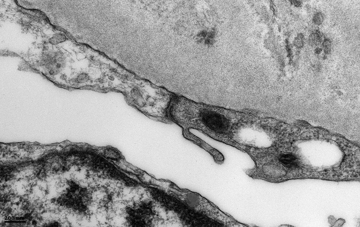
Are Langerhans histiocytes a normal component of the renal tubular epithelium? We have patient w uveitis + minimal inflammatory infiltrate on renal bx. EM shows structures that look like Birbeck granules (arrows). Could the presence of Langerhans cells indicate anything?#askrenal

Are Langerhans histiocytes a normal component of the renal tubular epithelium? We have patient w uveitis + minimal inflammatory infiltrate on renal bx. EM shows structures that look like Birbeck granules (arrows). Could the presence of Langerhans cells indicate anything?#askrenal
John Brealey (@johnbrealey) 's Twitter Profile Photo

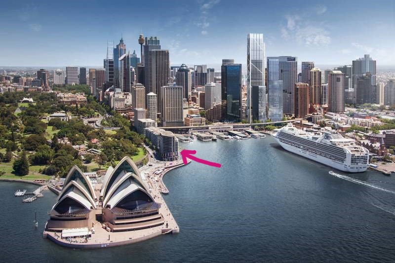
Last chance to register for the Electron Microscopy Tutorial Day & UltraPath XXI Conference, Sep 30-Oct 4. You can't beat the location. Follow the pink arrow and you'll find us discussing ultrastructural pathology at Circular Quay in the heart of Sydney. ultrapath.org

Last chance to register for the Electron Microscopy Tutorial Day &amp; UltraPath XXI Conference, Sep 30-Oct 4. You can't beat the location. Follow the pink arrow and you'll find us discussing ultrastructural pathology at Circular Quay in the heart of Sydney.
ultrapath.org
John Brealey (@johnbrealey) 's Twitter Profile Photo

Native kidney bx of a 50M w psoriasis. @electronmicroscopy shows membranous nephropathy (MN). Is there a relationship between psoriasis and MN? Could there be a PLA2 receptor auto-antibody that attacks both keratinocytes in skin + podocytes in glomeruli? Sanjeev Sethi #askrenal
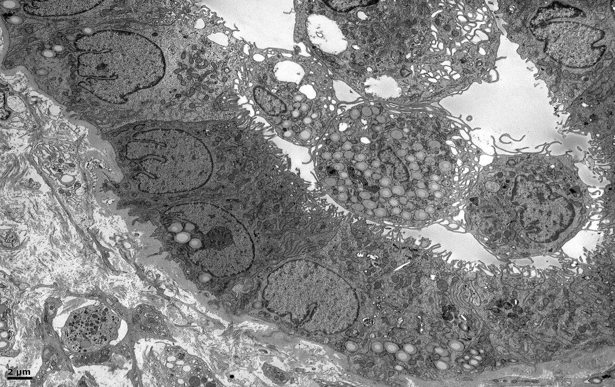
Native kidney bx of a 50M w psoriasis. @electronmicroscopy shows membranous nephropathy (MN). Is there a relationship between psoriasis and MN? Could there be a PLA2 receptor auto-antibody that attacks both keratinocytes in skin + podocytes in glomeruli? <a href="/SethiRenalPath/">Sanjeev Sethi</a> #askrenal
John Brealey (@johnbrealey) 's Twitter Profile Photo

Patient w membranous nephropathy. #electronmicroscopy shows electron-dense deposits (arrow) lining Bowman's capsule (BC) at the vascular pole, but deposits are not evident further along BC. Presumably the transition point between #podocyte and parietal epithelial cell phenotypes.

Patient w membranous nephropathy. #electronmicroscopy shows electron-dense deposits (arrow) lining Bowman's capsule (BC) at the vascular pole, but deposits are not evident further along BC. Presumably the transition point between #podocyte and parietal epithelial cell phenotypes.
John Brealey (@johnbrealey) 's Twitter Profile Photo

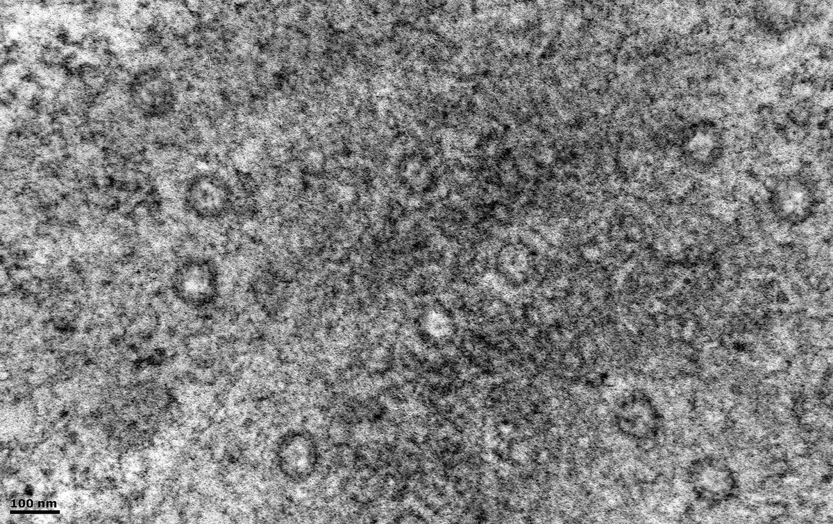
Kidney Bx. Looking for an infective agent by #electronmicroscopy. Found these structures that can be misinterpreted as viruses. They are nuclear pores. The plane of section has tangentially traversed the edge of a nucleus.

Kidney Bx. Looking for an infective agent by #electronmicroscopy.
Found these structures that can be misinterpreted as viruses. They are nuclear pores. The plane of section has tangentially traversed the edge of a nucleus.
John Brealey (@johnbrealey) 's Twitter Profile Photo

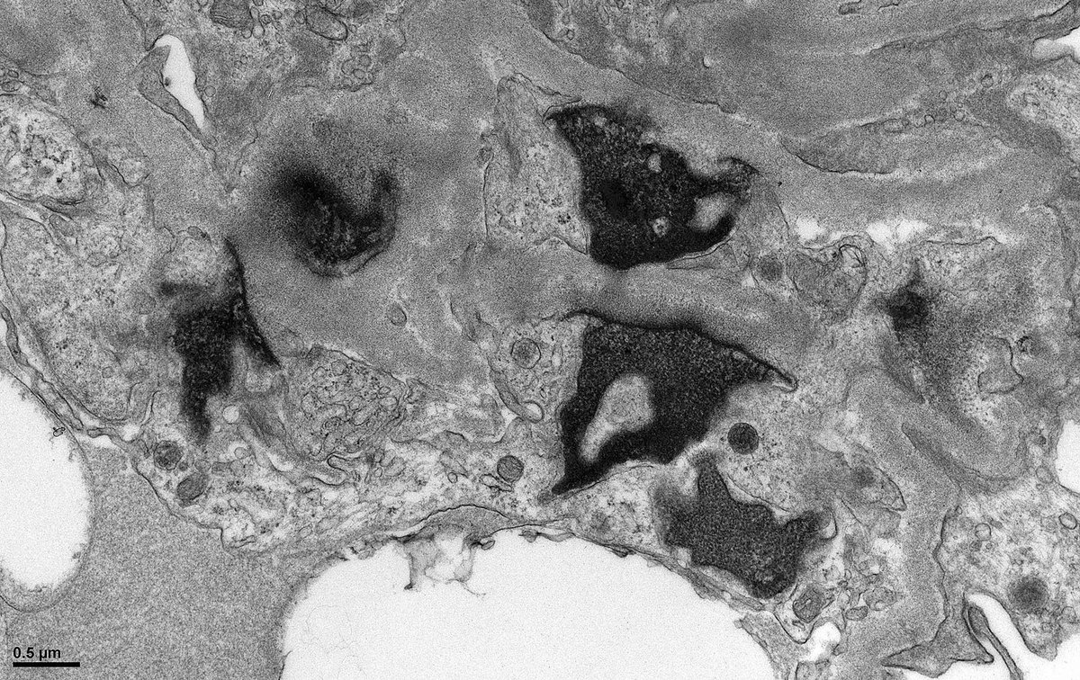
Native Kidney Bx #electronmicroscopy. #askrenal Any thoughts on these electron-dense structures? I interpret them as karyorrhectic debris. I see them rarely but have never seen them documented in an illustration. Interestingly this patient has 1+ C1q in capillary walls.

Native Kidney Bx #electronmicroscopy. #askrenal Any thoughts on these electron-dense structures? I interpret them as karyorrhectic debris. I see them rarely but have never seen them documented in an illustration. Interestingly this patient has 1+ C1q in capillary walls.
John Brealey (@johnbrealey) 's Twitter Profile Photo

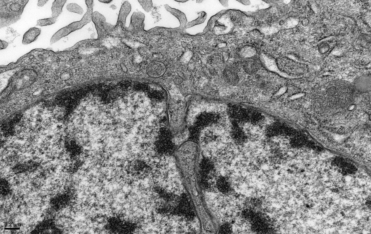
#electronmicroscopy Kidney bx shows a membranous nephropathy. #podocytes, typically post-mitotic, appear to be trying to divide. Nuclei are bi-lobed with narrow chromatin bridges. Cells are rounded with blebs + detachment from the GBM. Possibly an example of mitotic catastrophe.

#electronmicroscopy Kidney bx shows a membranous nephropathy. #podocytes, typically post-mitotic, appear to be trying to divide. Nuclei are bi-lobed with narrow chromatin bridges. Cells are rounded with blebs + detachment from the GBM. Possibly an example of mitotic catastrophe.
John Brealey (@johnbrealey) 's Twitter Profile Photo

#askrenal Can a plasmacytoma produce anti-LCAT autoantibodies? We have a case of a patient with a plasmacytoma. However, by #electronmicroscopy, the glomerular ultrastructure suggests LCAT deficiency.