MSS India (@msksocietyindia) 's Twitter Profile
MSS India

@msksocietyindia

The Musculoskeletal Society, India is a nonprofit association of Musculoskeletal Radiologists dedicated to teaching & spreading awareness about MSK Radiology

ID: 1831724875947384832

linkhttp://www.indianmss.org calendar_today05-09-2024 16:03:52

259 Tweet

1,1K Followers

129 Following

Sameer Raniga (@samrad77) 's Twitter Profile Photo

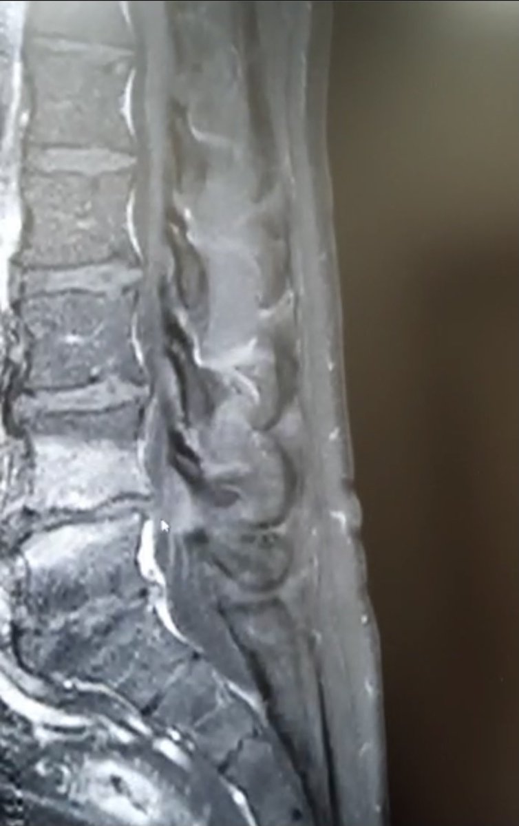
Young patient with lower backache. T2, T1, STIR and post-Gd Sagittal images- shows marrow edema involving the L4-5 vertebral end plates(L4>L5) with enhancement on Gd administration. L4-5 disc hyperintensity is also noted without enhancement. Modic type 1 or spondylodiskitis?

Young patient with lower backache. T2, T1, STIR and post-Gd Sagittal images- shows marrow edema involving the L4-5 vertebral end plates(L4>L5) with enhancement on Gd administration. L4-5 disc hyperintensity is also noted without enhancement. Modic type 1 or spondylodiskitis?
Sᴡɪss Uʟᴛʀᴀsᴏᴜɴᴅ Cᴇɴᴛᴇʀ - UZR (@rheumatology) 's Twitter Profile Photo

Compression neuropathy of the peroneal nerve secondary to a ganglion cyst Here some articles: pmc.ncbi.nlm.nih.gov/articles/PMC26… sciencedirect.com/science/articl… Here a case from last week:

Sameer Raniga (@samrad77) 's Twitter Profile Photo

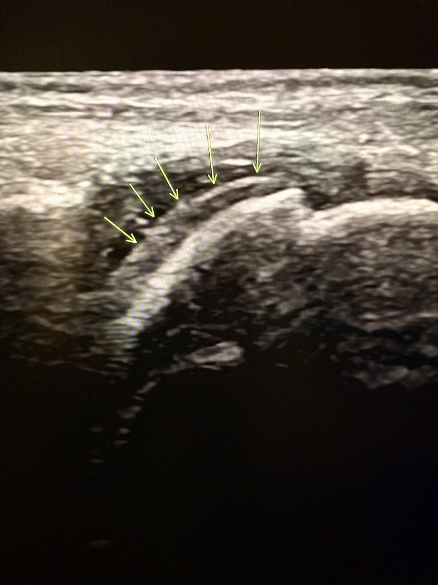
Double contour sign! It is a sonographic sign specific for gouty arthritis. Why does it occur? In gout, monosodium urate crystals deposit on the surface of the hyaline cartilage within the joint. This results in two distinct echogenic lines: 1.Deep line = normal

Double contour sign! 

It is a sonographic sign specific for gouty arthritis. 

Why does it occur?

In gout, monosodium urate crystals deposit on the surface of the hyaline cartilage within the joint. 

This results in two distinct echogenic lines:

1.Deep line = normal
MSS India (@msksocietyindia) 's Twitter Profile Photo

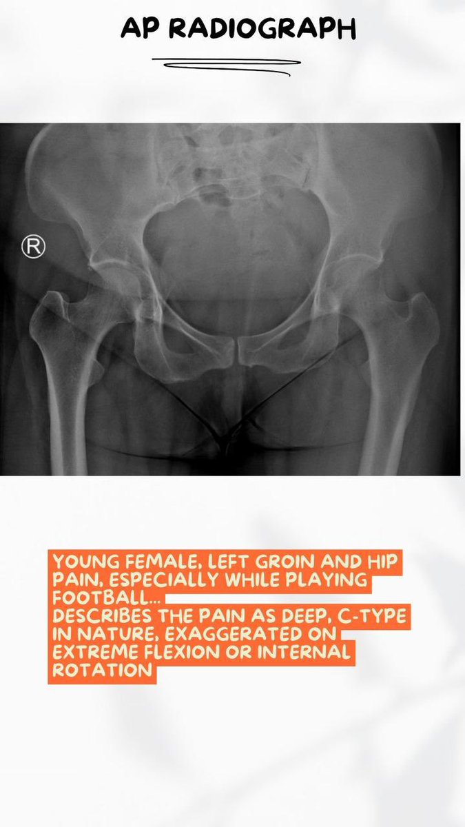
Femoroacetabular impingement (FAI) syndrome should only be applied to patients with a clinical disorder defined by the triad of: 1.Symptoms in young individuals 2.Clinical signs at physical examination 3.Characteristic radiological findings As a clinical radiologist, our focus

Femoroacetabular impingement (FAI) syndrome should only be applied to patients with a clinical disorder defined by the triad of:
1.Symptoms in young individuals
2.Clinical signs at physical examination
3.Characteristic radiological findings
As a clinical radiologist, our focus
MSS India (@msksocietyindia) 's Twitter Profile Photo

Let’s discuss the radiological signs of FAI … Cross-over sign - the superior portion of the anterior acetabular wall extends laterally beyond the posterior acetabular wall. •Posterior wall sign - the posterior wall projects medial to the center of the femoral head. •Ischial

Let’s discuss the radiological signs of FAI … Cross-over sign - the superior portion of the anterior acetabular wall extends laterally beyond the posterior acetabular wall.
•Posterior wall sign - the posterior wall projects medial to the center of the femoral head.
•Ischial
Suvinay Saxena (@drsuvi89) 's Twitter Profile Photo

There’s something humbling about handling a structure as fine and vital as a nerve. USG guided sural nerve biopsy, a small but significant procedure in the diagnostic workup of peripheral neuropathies. It’s a reminder that precision, patience, and respect for the human body.

MSS India (@msksocietyindia) 's Twitter Profile Photo

MSK RADIOLOGY FOR EVERYONE, Episode 2: Assessing lumbar radiographs post spine fixation. Many of us get scared when we come across a spine with hardware like fixators and interbody cages, but with a few tips and pointers we should be able to report them effectively. Hope this

MSK RADIOLOGY FOR EVERYONE, Episode 2: Assessing lumbar radiographs post spine fixation.
Many of us get scared when we come across a spine with hardware like fixators and interbody cages, but with a few tips and pointers we should be able to report them effectively. Hope this
MSS India (@msksocietyindia) 's Twitter Profile Photo

MSK RADIOLOGY FOR EVERYONE, continuing with Episode 2: Assessing lumbar radiographs post spine fixation. . Hope this helps! #msk #mskRad #FOAMRad #FOAMMed #radiology #RadRes #medicine #orthotwitter #MSKImaging #Radiology #Education #Musculoskeletal #FoamRad #medtwitter Sameer Raniga
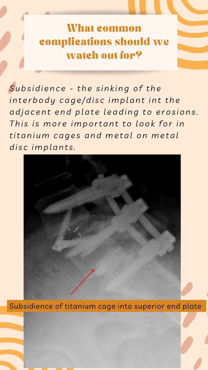
MSK RADIOLOGY FOR EVERYONE, continuing with Episode 2: Assessing lumbar radiographs post spine fixation.
. Hope this helps!

#msk #mskRad #FOAMRad #FOAMMed #radiology #RadRes #medicine #orthotwitter #MSKImaging #Radiology #Education #Musculoskeletal #FoamRad #medtwitter
<a href="/samrad77/">Sameer Raniga</a>
Sᴡɪss Uʟᴛʀᴀsᴏᴜɴᴅ Cᴇɴᴛᴇʀ - UZR (@rheumatology) 's Twitter Profile Photo

Uʟᴛʀᴀsᴏᴜɴᴅ ǫᴜɪᴢ Lᴇᴠᴇʟ: ᴍᴇᴅɪᴜᴍ Aɴᴛᴇʀɪᴏʀ sʜᴏᴜʟᴅᴇʀ 12 ᴡᴇᴇᴋs ᴀғᴛᴇʀ ʀᴇᴄᴏɴsᴛʀᴜᴄᴛɪᴏɴ ᴏғ ᴛʜᴇ sᴜʙsᴄᴀᴘᴜʟᴀʀɪs ᴛᴇɴᴅᴏɴ. Iᴛ ᴘɪɴᴄʜᴇs ᴅᴜʀɪɴɢ ɪɴᴛᴇʀɴᴀʟ ʀᴏᴛᴀᴛɪᴏɴ ᴀɴᴅ ᴛʜᴇʀᴇ ɪs ᴀ ᴘᴀɪɴғᴜʟ sᴛᴏᴘ. Wʜᴀᴛ ɪs ᴛʜᴇ ᴘʀᴏʙʟᴇᴍ?

sergio serrano belmar (@sserranobmsk) 's Twitter Profile Photo

Abdominal wall pain: differential diagnosis A 28-year-old soccer player presented with right periumbilical pain of 2 months' duration. An MRI was performed, which showed no abnormalities in the abdominal wall. He was diagnosed with anterior cutaneous nerve entrapment syndrome

Sameer Raniga (@samrad77) 's Twitter Profile Photo

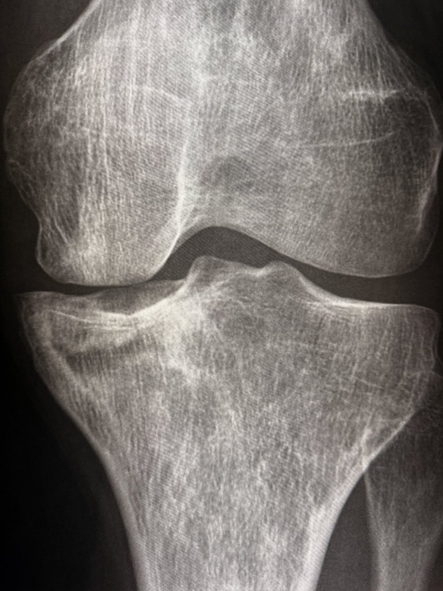
In sickle cell disease, chronic bone infarcts appear as intramedullary serpiginous sclerosis in the metadiaphyseal region with coarse, sclerotic trabecular pattern. Cortical thickening with “bone within bone” appearance. + AVN of distal femoral & proximal tibial condyles +

In sickle cell disease, chronic bone infarcts appear as intramedullary serpiginous sclerosis in the metadiaphyseal region with 
coarse, sclerotic trabecular pattern. Cortical thickening with “bone within bone” appearance. 

+ AVN of distal femoral &amp; proximal tibial condyles

+
Sameer Raniga (@samrad77) 's Twitter Profile Photo

Modern luxury is the ability to think clearly, sleep deeply, move slowly, and live quietly in a world designed to prevent all four. — Justin Welch

Sameer Raniga (@samrad77) 's Twitter Profile Photo

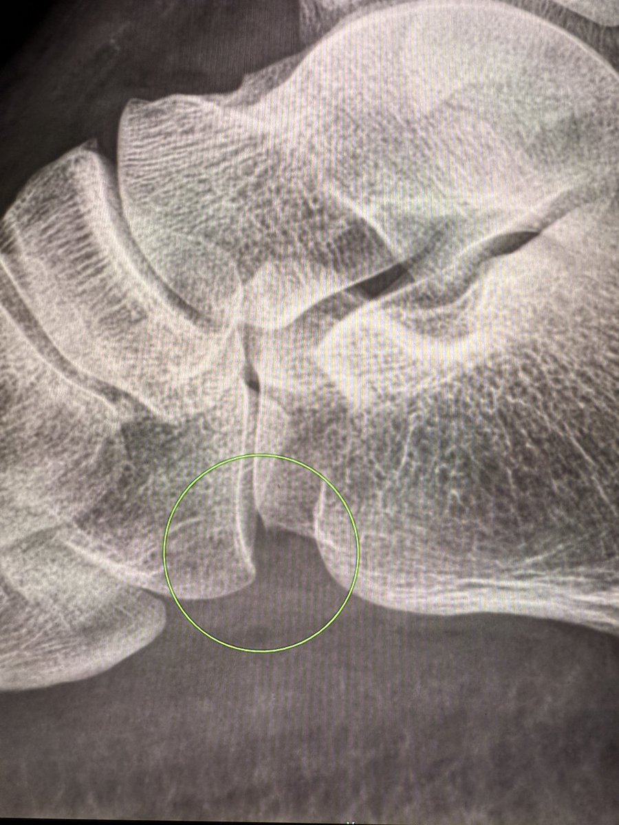
On lateral ankle radiographs, the cuboid often appears inferior to the calcaneum at the calcaneocuboid joint—up to one-fourth of the articular surface. It’s normal. Not every step-down is subluxation. —Pearls and pitfalls from my reporting list

On lateral ankle radiographs, the cuboid often appears inferior to the calcaneum at the calcaneocuboid joint—up to one-fourth of the articular surface.

It’s normal. Not every step-down is subluxation.

—Pearls and pitfalls from my reporting list
Sameer Raniga (@samrad77) 's Twitter Profile Photo

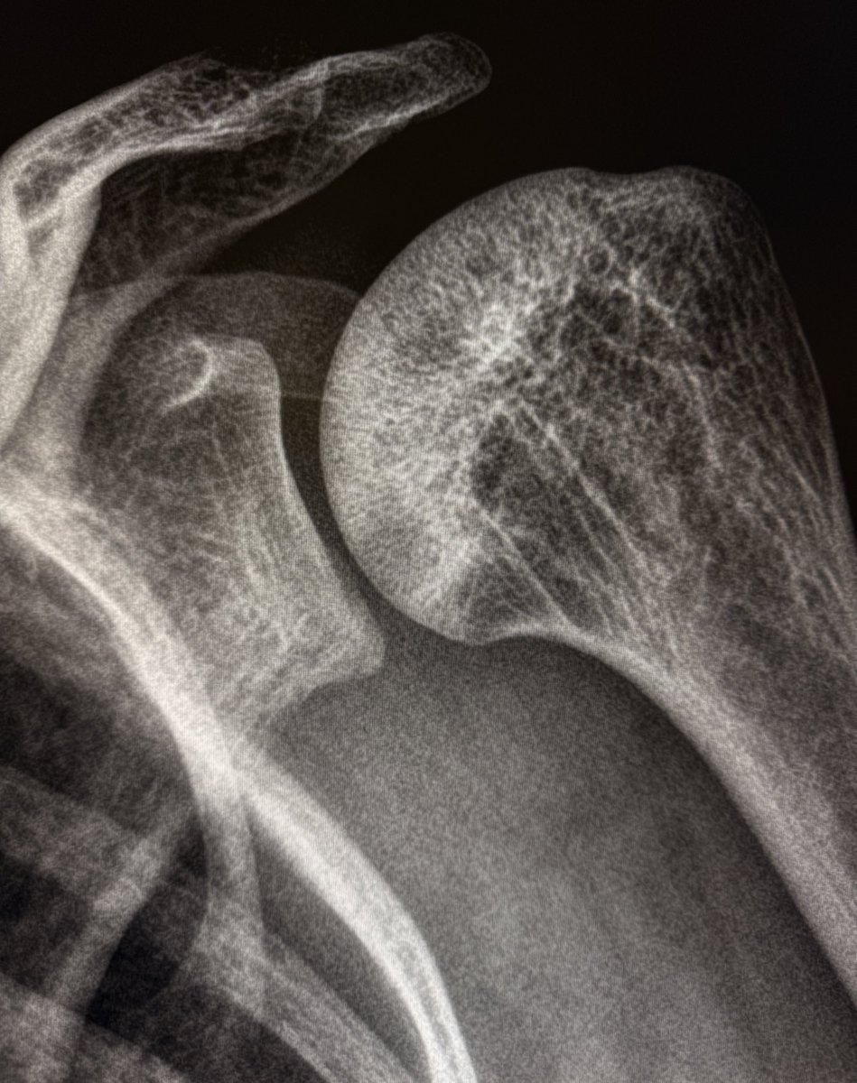
Nothing fancy today! This is just a nice example of the “Snowcap sign” of AVN of the humeral heads (bilateral) on radiographs. The sclerotic, dense necrotic bone beneath a preserved articular surface resembles a snow-covered mountain peak, a deceptive summit above a crumbling

Nothing fancy today! 

This is just a nice example of the “Snowcap sign” of AVN of the humeral heads (bilateral) on radiographs. 

The sclerotic, dense necrotic bone beneath a preserved articular surface resembles a snow-covered mountain peak, a deceptive summit above a crumbling
sergio serrano belmar (@sserranobmsk) 's Twitter Profile Photo

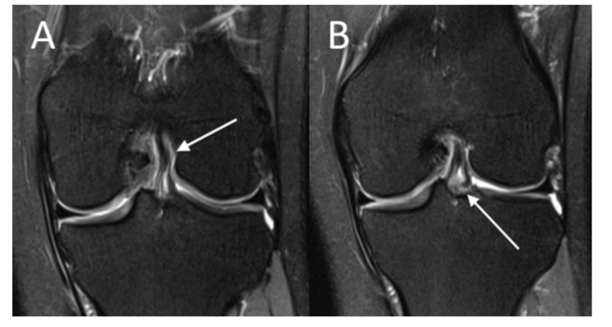
Management of Partial Tears of the Anterior Cruciate Ligament: A Review of the Anatomy, Diagnosis, and Treatment DOI: 10.5435/JAAOS-D-20-00242 This review article discusses partial anterior cruciate ligament (ACL) tears, aiming to provide clinicians with guidance on anatomy,

Management of Partial Tears of the Anterior Cruciate Ligament: A Review of the Anatomy, Diagnosis, and Treatment
DOI: 10.5435/JAAOS-D-20-00242

This review article discusses partial anterior cruciate ligament (ACL) tears, aiming to provide clinicians with guidance on anatomy,