Maëlle Saliba (@maellesaliba) 's Twitter Profile
Maëlle Saliba

@maellesaliba

Thoracic Pathology fellow at Memorial Sloan Kettering Cancer Center

ID: 1440902954

calendar_today19-05-2013 11:16:05

76 Tweet

103 Followers

98 Following

Jonathan Lai (@jonlaimd) 's Twitter Profile Photo

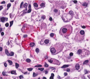
Sharing one of my favourite slides from autopsy case. Hemophagocytic lymphohistiocytosis: Macrophages behaving badly, devouring an innocent red blood cells (AKA Erythrophagocytosis). Overall mortality >60%. Ddx: Rosai-Dorfman Disease (lymphocytophagocytosis)

Sharing one of my favourite slides from autopsy case. Hemophagocytic lymphohistiocytosis: Macrophages behaving badly, devouring an innocent red blood cells (AKA Erythrophagocytosis). Overall mortality >60%. Ddx: Rosai-Dorfman Disease (lymphocytophagocytosis)
Xiaoyin "Sara" Jiang, MD (She/Her) (@sara_jiang) 's Twitter Profile Photo

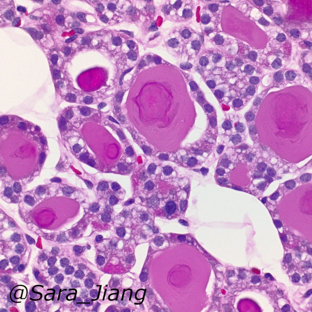
#PathPitfall, Frozens edition: Pseudofollicular change can occur in parathyroid (seen here) - easy to mistake for thyroid tissue on a small sample on frozens! Tips: if you see crystals in the "colloid", likely to be truly thyroid: pubmed.ncbi.nlm.nih.gov/24618617/

#PathPitfall, Frozens edition:
Pseudofollicular change can occur in parathyroid (seen here) - easy to mistake for thyroid tissue on a small sample on frozens! Tips: if you see crystals in the "colloid", likely to be truly thyroid: pubmed.ncbi.nlm.nih.gov/24618617/
Priya Rao, MD (@priyaraomd) 's Twitter Profile Photo

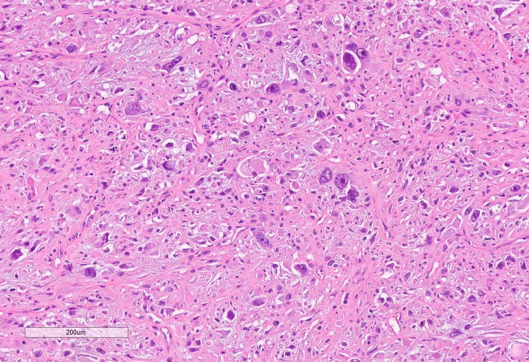
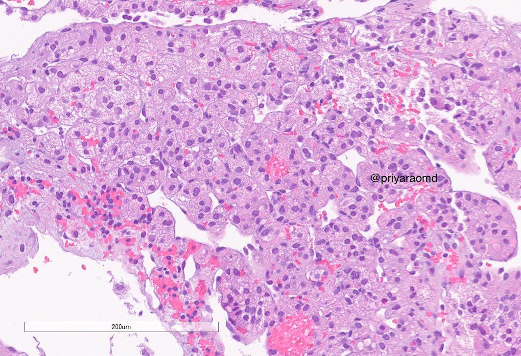
Two sections from within the same kidney tumor. I received a LN biopsy a few months later, that looks like the 2nd pic. Without the prior, it would have been difficult to classify solely on the HG component. Dx: Chromophobe RCC with sarcomatoid dedifferentiation. #GUPath

Two sections from within the same kidney tumor. I received a LN biopsy a few months later, that looks like the 2nd pic. Without the prior, it would have been difficult to classify solely on the HG component. 

Dx: Chromophobe RCC with sarcomatoid dedifferentiation. #GUPath
Meghana Agni MD (@meghanaagnimd) 's Twitter Profile Photo

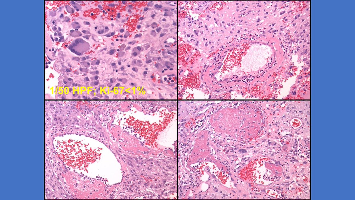
Just presented this to the Chicago Pathology Society! 51 yo M presented w/ painful mid-back lump that grew rapidly over a few weeks. He had a 30 lb weight loss over 3 mo, fatigue, and severe anemia. MRI: 5.6 x 4.3 x 3.6 cm paraspinal soft tissue mass. Your #DDx? University of Chicago Pathology #IRAP21
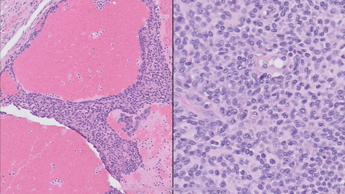
Just presented this to the 
<a href="/ChicagoPathSoc/">Chicago Pathology Society</a>!

51 yo M presented w/ painful mid-back lump that grew rapidly over a few weeks. He had a 30 lb weight loss over 3 mo, fatigue, and severe anemia. MRI: 5.6 x 4.3 x 3.6 cm paraspinal soft tissue mass. Your #DDx? <a href="/UChicagoPath/">University of Chicago Pathology</a> #IRAP21
Albert Alhatem (@aalhatem_md) 's Twitter Profile Photo

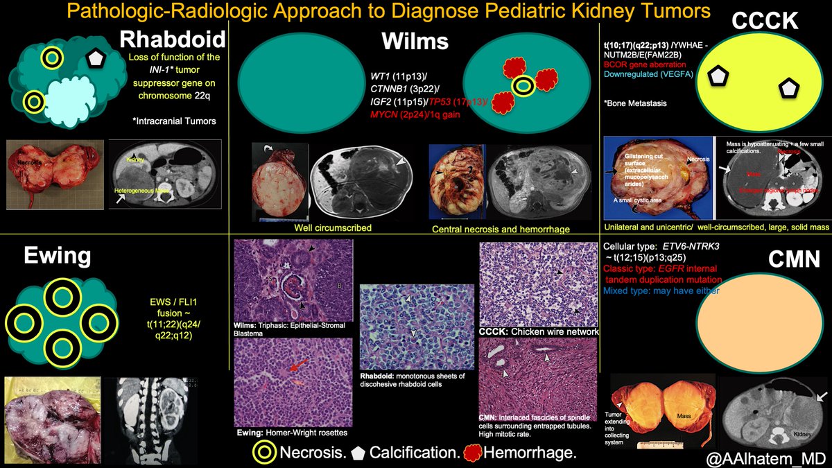
Giving a presentation to our residents, so I thought to share with #TwitterFamily the pathologic-radiologic approach to the diagnosis of pediatric kidney tumors & some free points on the #boardexams #pathresidents. Pics source: Chung et al 2016 radiographics.rsna.org.

Giving a presentation to our residents, so I thought to share with #TwitterFamily the pathologic-radiologic approach to the diagnosis of pediatric kidney tumors &amp; some free points on the #boardexams #pathresidents. Pics source: Chung et al 2016 radiographics.rsna.org.
Johns Hopkins Pathology (@jhupath) 's Twitter Profile Photo

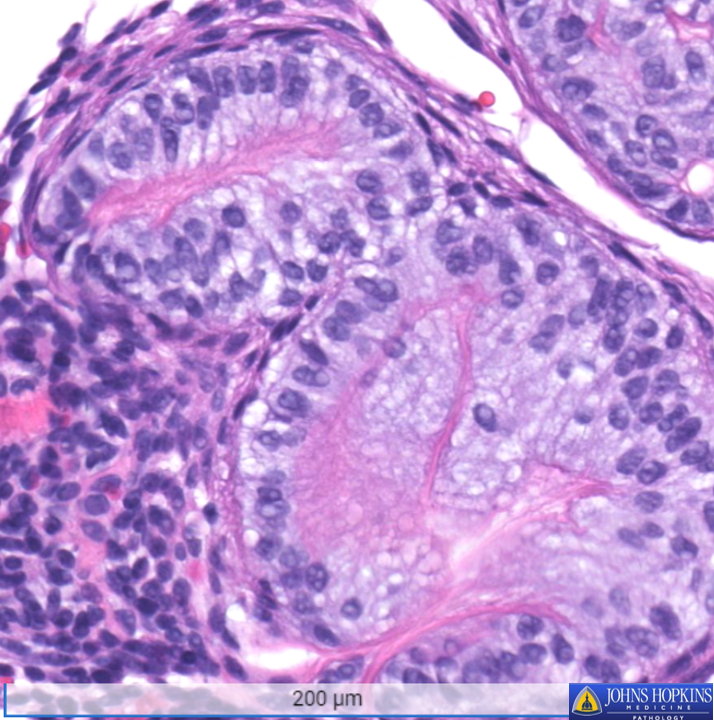
A teenage patient presents with a mandible cyst, shown below. What is your diagnosis? Answer and explanation to follow! #Pathology #MedEd

A teenage patient presents with a mandible cyst, shown below. What is your diagnosis? Answer and explanation to follow! #Pathology #MedEd
Priya Rao, MD (@priyaraomd) 's Twitter Profile Photo

I *still* have to remind my fellows to order both a panker/EMA & PAX8 in suspected cases of MIT RCCs. PAX8 expression correlates well with TFE3 staining on IHC. Look for TFE3 expression in areas that have strong PAX8 staining. Weak or neg PAX8 may be due to poor tumor fixation.

Samson W. Fine, MD (@rovingatuscap) 's Twitter Profile Photo

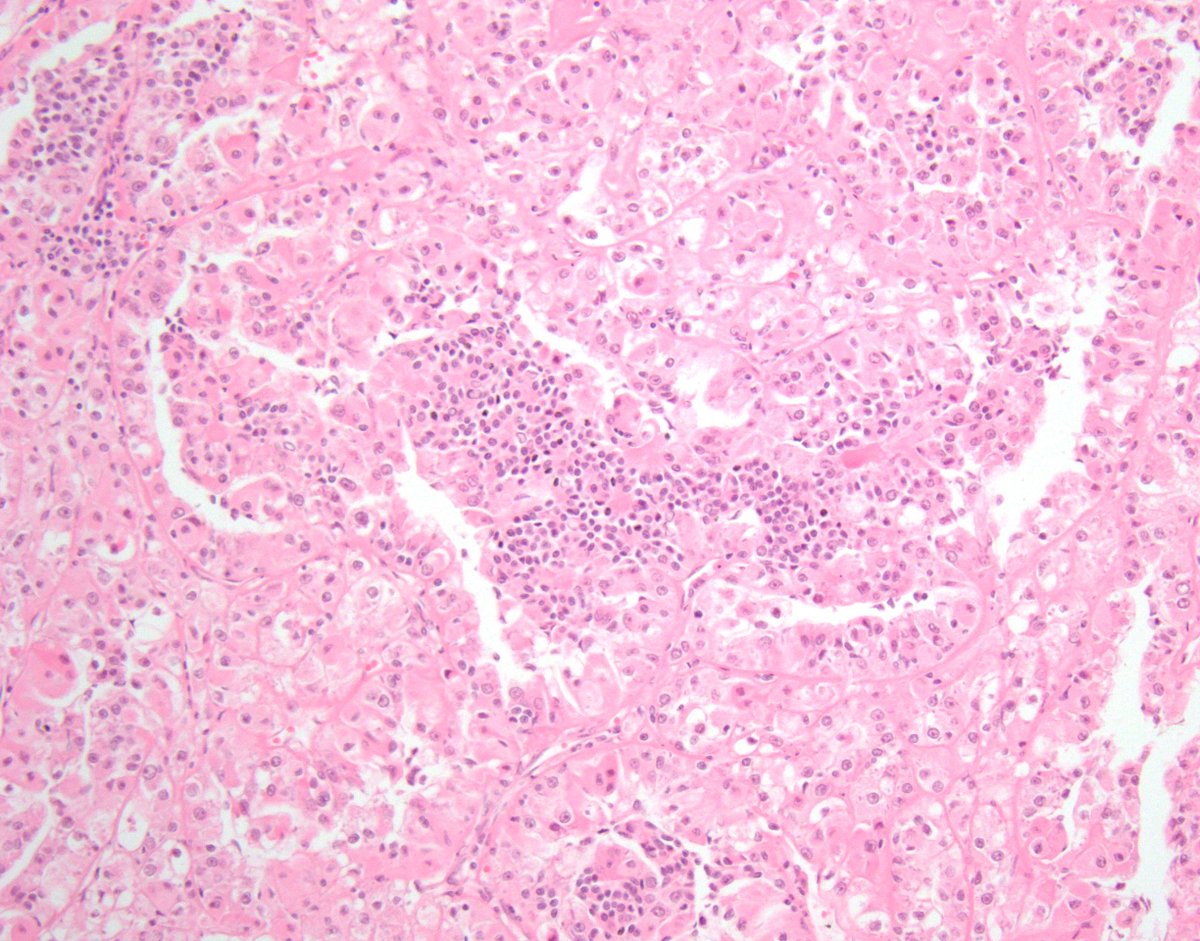
MITF Family Translocation-Associated RCC #2 TFEB (6p21) Rearranged 🔬biphasic morphology: - larger epithelioid cells w/clear to eosinophilic cytoplasm - smaller cells clustered around nodules of basement membrane material #gupath #kidneycancer #KidneyCancerAwarenessMonth 1/

MITF Family Translocation-Associated RCC #2

TFEB (6p21) Rearranged 

🔬biphasic morphology: 
- larger epithelioid cells w/clear to eosinophilic cytoplasm
- smaller cells clustered around nodules of basement membrane material

#gupath #kidneycancer #KidneyCancerAwarenessMonth 1/
WebPathology (@webpathology) 's Twitter Profile Photo

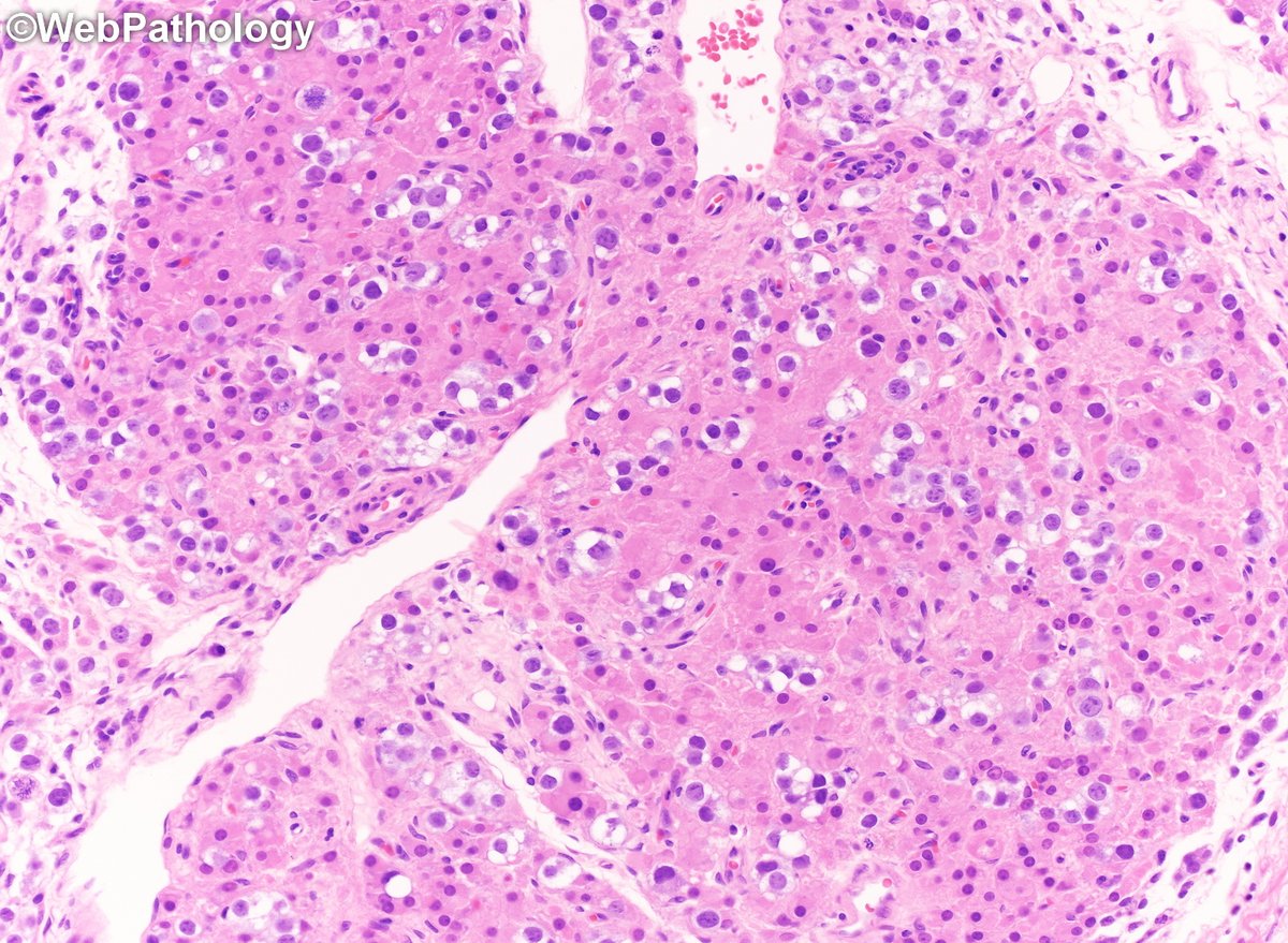
Common tumor with an uncommon presentation. Testicular tumor presenting with gynecomastia. What's going on here? Labs. and additional images to follow. #GUPath

Common tumor with an uncommon presentation. Testicular tumor presenting with gynecomastia. What's going on here? Labs. and additional images to follow. #GUPath
Natasha Rekhtman MD PhD (@natasharekhtman) 's Twitter Profile Photo

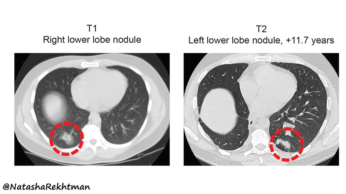
1/ Excited to share the latest #PulmPath study from our group. Let me introduce it with a case. This patient had a complete resection of lung adenocarcinoma. 11+ yrs later, he has a new contralateral tumor. No nodal or distant metastases.

1/ Excited to share the latest #PulmPath study from our group. Let me introduce it with a case. 

This patient had a complete resection of lung adenocarcinoma. 11+ yrs later, he has a new contralateral tumor. No nodal or distant metastases.
Maëlle Saliba (@maellesaliba) 's Twitter Profile Photo

Honored and grateful to receive the ADASP surgical pathology award for our abstract on IMA. Thank you to my mentors at Columbia Pathology for making this possible! #USCAP2023
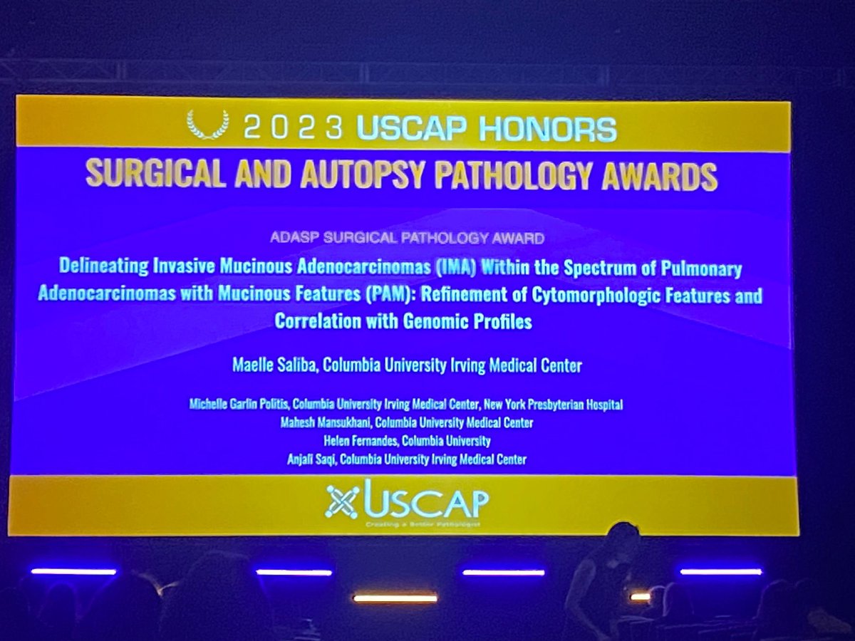
Honored and grateful to receive the ADASP surgical pathology award for our abstract on IMA. Thank you to my mentors at <a href="/ColumbiaPath/">Columbia Pathology</a> for making this possible! #USCAP2023
Papanicolaou Society (@papsociety) 's Twitter Profile Photo

Case of the month : A 72Y/O woman underwent an endobronchial #USG #FNA of a 1.0 cm well-circumscribed mass. What is the best diagnosis? submitted by: Maëlle Saliba Michelle A. Garlin Politis Niyati Desai and Dr.Saqi! #papcyto click 👉 for ans: papsociety.com/cotm22/cotmdec…

Case of the month : A 72Y/O woman underwent an endobronchial #USG #FNA of a 1.0 cm well-circumscribed mass. What is the best diagnosis? submitted by: <a href="/MaelleSaliba/">Maëlle Saliba</a> <a href="/MichelleGarlin/">Michelle A. Garlin Politis</a> <a href="/NiyatiDesai13/">Niyati Desai</a> and Dr.Saqi! #papcyto click 👉 for ans: papsociety.com/cotm22/cotmdec…