Majid Ahsan (@majid_ahsan) 's Twitter Profile
Majid Ahsan

@majid_ahsan

ID: 214634476

calendar_today11-11-2010 21:39:46

44 Tweet

40 Followers

68 Following

European Society of Cardiology (@escardio) 's Twitter Profile Photo

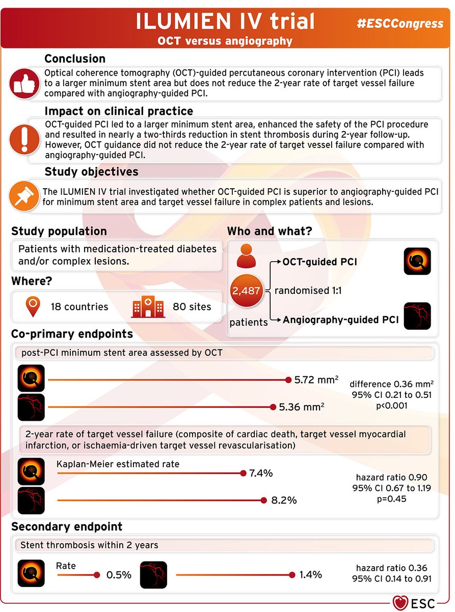
Optical coherence tomography (OCT)-guided percutaneous coronary intervention (PCI) leads to a larger minimum stent area but does not reduce the two-year rate of target vessel failure compared with angiography-guided #PCI ⬇️ #ESCCongress ILUMIEN IV trial

Optical coherence tomography (OCT)-guided  percutaneous coronary intervention (PCI) leads to a larger minimum stent area but does not reduce  the two-year rate of target vessel failure compared with angiography-guided #PCI ⬇️

#ESCCongress ILUMIEN IV trial
Deutsche Gesellschaft für Kardiologie (@dgk_org) 's Twitter Profile Photo

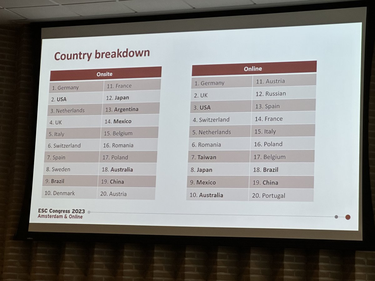
Teilnehmerzahlen vom #ESCCongress nach Ländern. Aus keinem anderen Land waren mehr Kardiologinnen und Kardiologen vor Ort als aus Deutschland. Onsite und online. 🙂👏

Teilnehmerzahlen vom #ESCCongress nach Ländern. 

Aus keinem anderen Land waren mehr Kardiologinnen und Kardiologen vor Ort als aus Deutschland. Onsite und online. 🙂👏
Majid Ahsan (@majid_ahsan) 's Twitter Profile Photo

Pre-op echo images of a 74-year-old patient with chronic Stanford type A (DeBakey II) aortic dissection. Clear visualization of the dissection membrane

Majid Ahsan (@majid_ahsan) 's Twitter Profile Photo

80-year-old patient with a history of TAVR in 2023, now presenting with endocarditis of the TAVR prosthesis. #Echo #Cardiology #TAVR

Majid Ahsan (@majid_ahsan) 's Twitter Profile Photo

60-year-old patient with a perimembranous VSD due to biopatch dehiscence after aortic root and valve replacement for florid endocarditis. #Echo #VSD #Cardiology #Endocarditis

Majid Ahsan (@majid_ahsan) 's Twitter Profile Photo

77-year-old patient with prior mitral valve repair (2017) and MitraClip (2020). Now presenting with severe residual mitral regurgitation: •Paravalvular jet due to ring dehiscence •Valvular jet due to AML lifting, with near detachment of the clip #Echo #Cardiology

Majid Ahsan (@majid_ahsan) 's Twitter Profile Photo

66-year-old patient with severe primary mitral regurgitation due to flail of the posterior mitral leaflet (P2 segment) #Echo #Cardiology #Mitral

Majid Ahsan (@majid_ahsan) 's Twitter Profile Photo

83-year-old patient with infective endocarditis after TAVR. Echo shows a perforated abscess cavity in the mitral-aortic intervalvular fibrosa, extending into the anterior mitral leaflet, causing severe mitral regurgitation. #Echo #Cardiology #Endocarditis

Majid Ahsan (@majid_ahsan) 's Twitter Profile Photo

67-year-old patient with severe mitral stenosis, likely due to prior rheumatic fever. Echo shows thickened, doming AML with commissural fusion of both leaflets — classic rheumatic mitral valve disease. #Echo #Cardiology #MitralStenosis

Majid Ahsan (@majid_ahsan) 's Twitter Profile Photo

61-year-old patient with a large aneurysm of the basomedial posterior wall and medioapical inferoseptal wall, complicated by an apical inferoseptal VSD (post-infarction). 🔎 Echo shows a clear left-to-right shunt through the infarct-related septal defect. #Echo #VSD