Tatyana Milman (@milmantatyana) 's Twitter Profile
Tatyana Milman

@milmantatyana

Board certified ophthalmologist and anatomic pathologist. Practice eye pathology and comprehensive ophthalmology at Wills Eye Hospital.

ID: 1108846993596514304

calendar_today21-03-2019 21:44:59

179 Tweet

312 Followers

53 Following

Tatyana Milman (@milmantatyana) 's Twitter Profile Photo

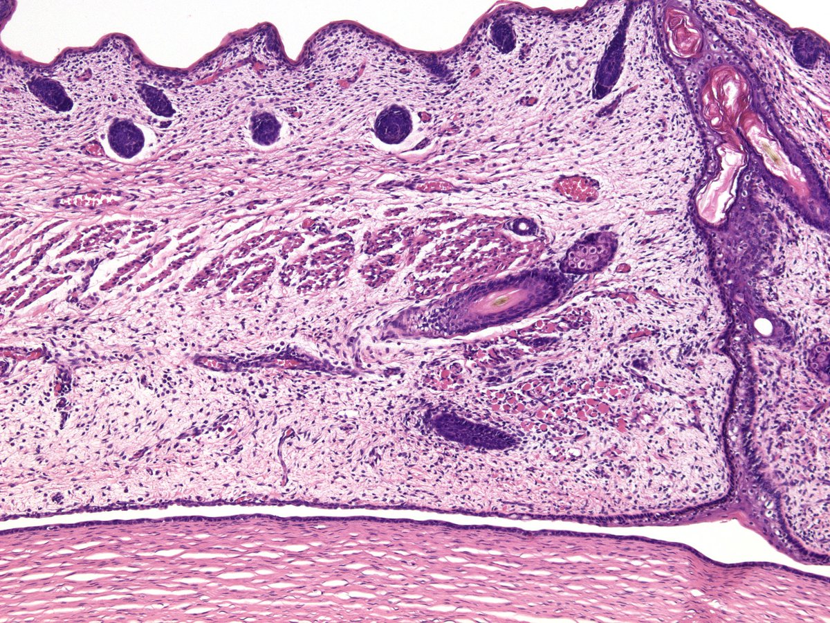
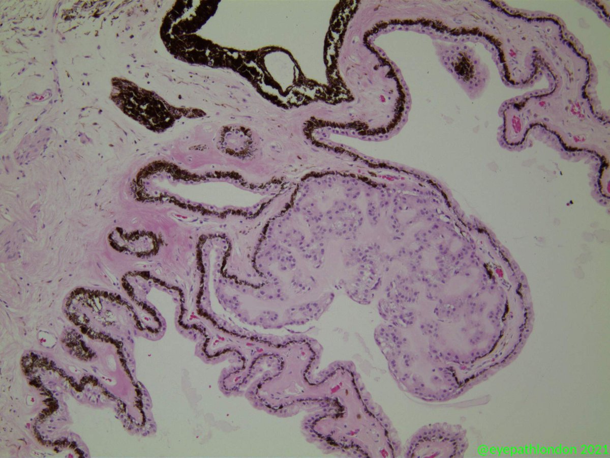
18 week gestational age fetus. The eyelid development with early adnexal appendages, early Meibomian gland ducts. The upper and lower eyelids are still fused together, just about ready to separate. Note the absence of Bowman layer in the underlying developing cornea.

18 week gestational age fetus. The eyelid development with early adnexal appendages, early Meibomian gland ducts. The upper and lower eyelids are still fused together, just about ready to separate. Note the absence of Bowman layer in the underlying developing cornea.
Tatyana Milman (@milmantatyana) 's Twitter Profile Photo

Our fantastic resident Dr Lucy Cobbs presenting a case of metastatic cutaneous melanoma to the eye to our visiting professor Dr Dryja from MEEI. Photos taken by our chief resident Dr Kalla Gervasio.

Our fantastic resident Dr Lucy Cobbs presenting a case of metastatic cutaneous melanoma to the eye to our visiting professor Dr Dryja from MEEI. Photos taken by our chief resident Dr Kalla Gervasio.
Tatyana Milman (@milmantatyana) 's Twitter Profile Photo

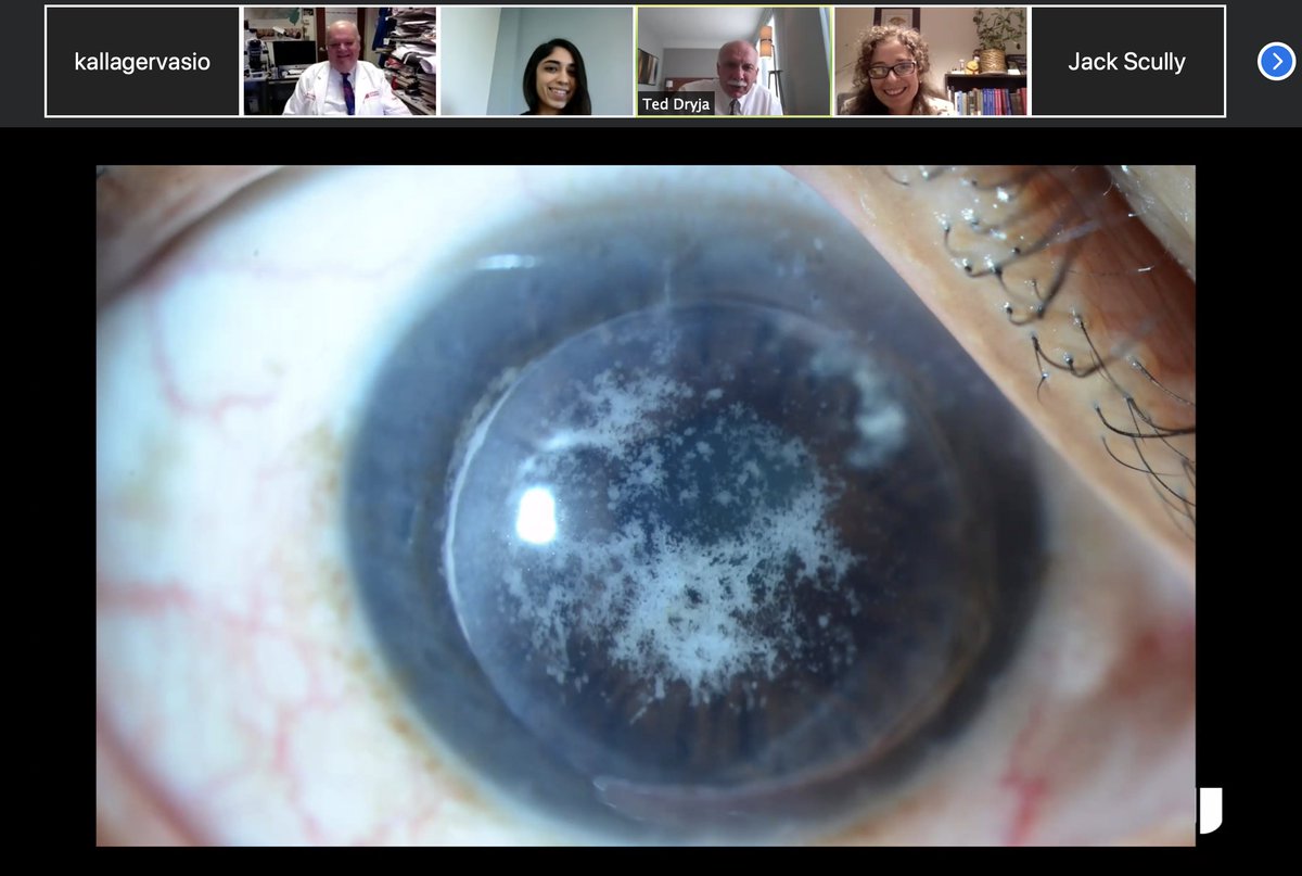
And here's another fantastic Wills Eye resident Rakhi presenting on granular dystrophy recurring at DALK graft-recipient interface.

And here's another fantastic Wills Eye resident Rakhi presenting on granular dystrophy recurring at DALK graft-recipient interface.
Tatyana Milman (@milmantatyana) 's Twitter Profile Photo

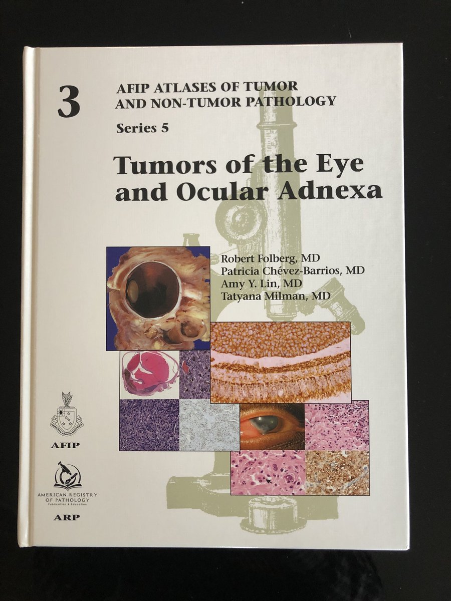
My coauthors and I hope that this book will help pathologists to guide ocular oncologists to preserve life and sight. Details on availability to follow …

My coauthors and I hope that this book will help pathologists to guide ocular oncologists to preserve life and sight. Details on availability to follow …
JAMA Ophthalmology (@jamaophth) 's Twitter Profile Photo

A 77-year-old presents with blurred vision in the left eye secondary to a corneal lesion. What would you do next? ja.ma/2WJyqL2

ARP Press (@arp_press) 's Twitter Profile Photo

Starting next week, we’ll feature beautiful images from our Series V, Number 3: Tumors of the Eye and #Ocular Adnexa by Robert Folberg @patychevez1 Amy Lin, MD and Tatyana Milman. Fascicle available on arppress.org. #eyetumor #eyepath #pathology #tumor #pathresidents
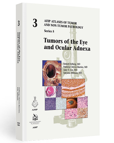
Starting next week, we’ll feature beautiful images from our Series V, Number 3: Tumors of the Eye and #Ocular Adnexa by <a href="/EyePath/">Robert Folberg</a> @patychevez1 <a href="/StudyPathology/">Amy Lin, MD</a> and <a href="/MilmanTatyana/">Tatyana Milman</a>.

Fascicle available on arppress.org.

#eyetumor #eyepath #pathology #tumor #pathresidents
Charles Eberhart (@neuroeyepath) 's Twitter Profile Photo

A brown #cornea due to copper deposition in Descemets and endothelium in a patient with #MGUS. Special stain for copper upper right, and IgG (faint staining) lower right. #eyepath

A brown #cornea due to copper deposition in Descemets and endothelium in a patient with #MGUS. Special stain for copper upper right, and IgG (faint staining) lower right. #eyepath
ARP Press (@arp_press) 's Twitter Profile Photo

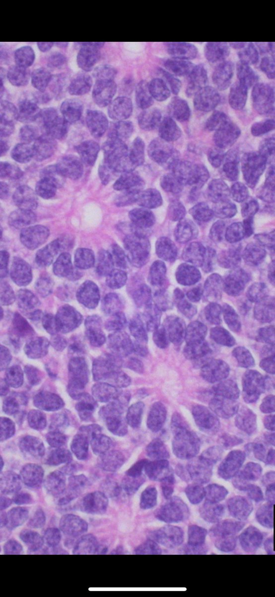
Featured Images Set #2: Tumors of the Eye and Ocular Adnexa by Dr. Folberg Robert Folberg @patychevez1 Amy Lin, MD & Tatyana Milman Differentiation in retinoblastoma (figure 9-15 A). Visit arppress.org for book info. #eyetumor #eyepath #pathology #tumor #pathresidents
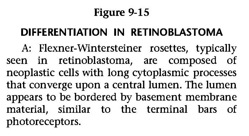
Featured Images Set #2: Tumors of the Eye and Ocular Adnexa by Dr. Folberg <a href="/EyePath/">Robert Folberg</a> @patychevez1 <a href="/StudyPathology/">Amy Lin, MD</a> &amp; <a href="/MilmanTatyana/">Tatyana Milman</a>

Differentiation in retinoblastoma (figure 9-15 A).

Visit arppress.org for book info.

#eyetumor #eyepath #pathology #tumor #pathresidents
ARP Press (@arp_press) 's Twitter Profile Photo

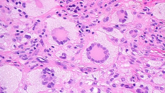
Featured Images Set #5: Tumors of the Eye and Ocular Adnexa by Dr. Folberg Robert Folberg @patychevez1 Amy Lin, MD & Tatyana Milman. Retinoblastoma: cytopathologic features (figure 9-17). Order book at arppress.org. #eyetumor #eyepath #pathology #tumor #pathresidents
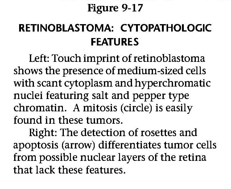
Featured Images Set #5: Tumors of the Eye and Ocular Adnexa by Dr. Folberg <a href="/EyePath/">Robert Folberg</a> @patychevez1 <a href="/StudyPathology/">Amy Lin, MD</a> &amp; <a href="/MilmanTatyana/">Tatyana Milman</a>.

Retinoblastoma: cytopathologic features (figure 9-17).
 
Order book at arppress.org.

#eyetumor #eyepath #pathology #tumor #pathresidents
ARP Press (@arp_press) 's Twitter Profile Photo

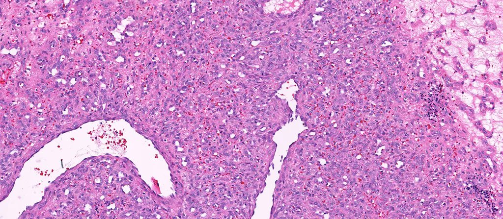
Featured Images Set #6: Tumors of the Eye and Ocular Adnexa by Dr. Folberg Robert Folberg @patychevez1 Amy Lin, MD & Tatyana Milman Retinoblastoma: immunohistochemistry (fig 9-21A). Visit arppress.org for book info. #eyetumor #eyepath #pathology #tumor #pathresidents
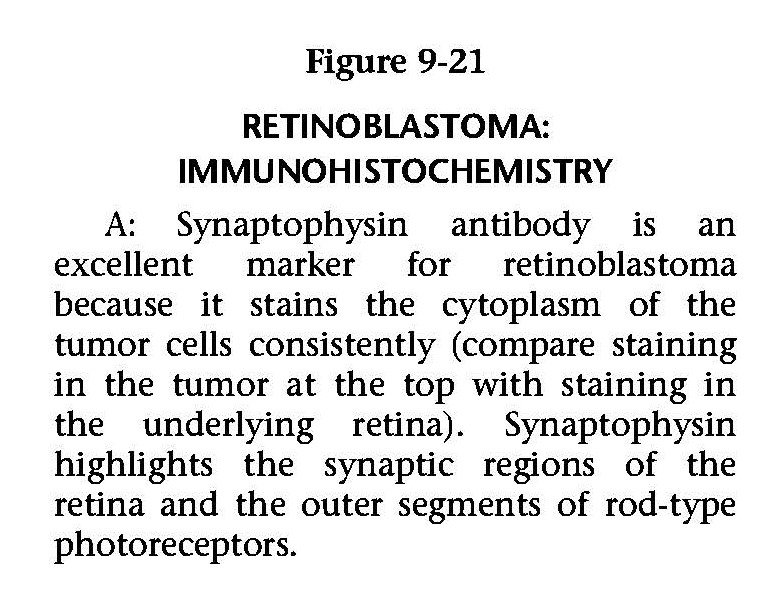
Featured Images Set #6: Tumors of the Eye and Ocular Adnexa by Dr. Folberg <a href="/EyePath/">Robert Folberg</a> @patychevez1 <a href="/StudyPathology/">Amy Lin, MD</a> &amp; <a href="/MilmanTatyana/">Tatyana Milman</a>

Retinoblastoma: immunohistochemistry (fig 9-21A). 

Visit arppress.org for book info.

#eyetumor #eyepath #pathology #tumor #pathresidents
Jerad Gardner, MD (@jmgardnermd) 's Twitter Profile Photo

Eye tumor from a baby. Entire globe of eye cut in half front to back. Amazing pics by Dr. Monica Evans. Your diagnosis? Reply then click the link to check your answer kikoxp.com/posts/4321 #pathologists #pathology #pathTwitter #ophthalmology #ophtho #eyepath

Eye tumor from a baby. Entire globe of eye cut in half front to back. Amazing pics by Dr. Monica Evans. Your diagnosis? Reply then click the link to check your answer kikoxp.com/posts/4321 #pathologists #pathology #pathTwitter #ophthalmology #ophtho #eyepath
Jerad Gardner, MD (@jmgardnermd) 's Twitter Profile Photo

Skin Adnexal tumor quiz. View these 12 unknown digital slides via pathpresenter.com: pathpresenter.net/#/public/prese…. Quiz yourself, then check your answers here: kikoxp.com/posts/3824 #dermpath #dermatology #dermatologia #dermtwitter #pathologists #pathology #pathTwitter
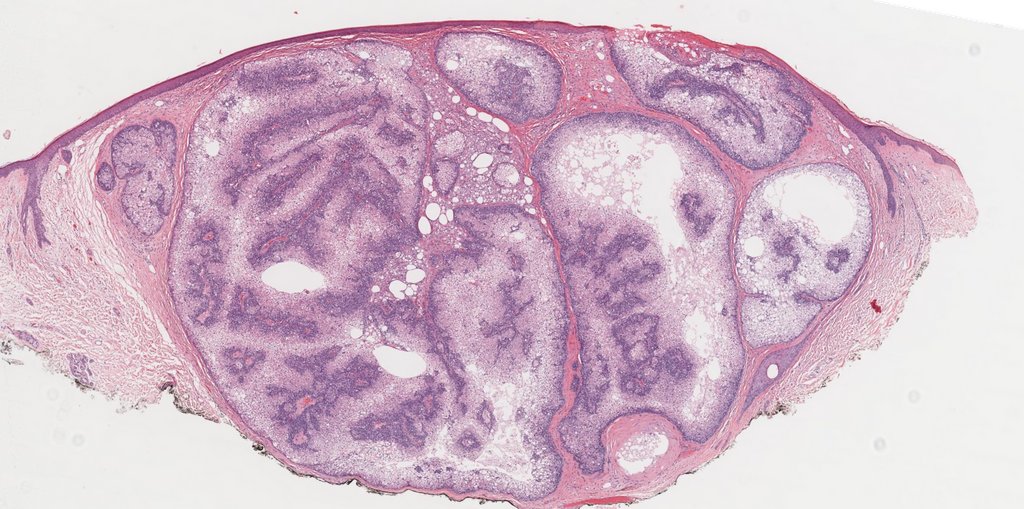
Skin Adnexal tumor quiz. View these 12 unknown digital slides via <a href="/pathpresenter/">pathpresenter.com</a>: pathpresenter.net/#/public/prese…. Quiz yourself, then check your answers here: kikoxp.com/posts/3824 #dermpath #dermatology #dermatologia #dermtwitter #pathologists #pathology #pathTwitter
Jerad Gardner, MD (@jmgardnermd) 's Twitter Profile Photo

Yellow-red papule. Forehead of 5yo boy. One pic only. Your diagnosis? Answer & 5 minute video here ➡️ kikoxp.com/posts/6846. #pathology #pathologists #pathTwitter #dermpath #dermatology #dermatologia #dermtwitter

Yellow-red papule. Forehead of 5yo boy. One pic only. Your diagnosis? Answer &amp; 5 minute video here ➡️ kikoxp.com/posts/6846. #pathology #pathologists #pathTwitter #dermpath #dermatology #dermatologia #dermtwitter
Jerad Gardner, MD (@jmgardnermd) 's Twitter Profile Photo

Eye enucleation from an adult. Your diagnosis? Where does it usually metastasize first? Answers: kikoxp.com/posts/5912. Amazing pics by Dr. Monica Evans. #pathologists #pathology #pathTwitter #grosspath #Ophthotwitter #ophtho #MedStudentTwitter #medtwitter Dr. Glaucomflecken

Eye enucleation from an adult. Your diagnosis? Where does it usually metastasize first? Answers: kikoxp.com/posts/5912. Amazing pics by Dr. Monica Evans. #pathologists #pathology #pathTwitter #grosspath #Ophthotwitter #ophtho #MedStudentTwitter #medtwitter <a href="/DGlaucomflecken/">Dr. Glaucomflecken</a>
Christopher Heaphy (@chris_heaphy) 's Twitter Profile Photo

Latest collaborative work with Wills Eye Hospital Johns Hopkins Pathology confirms frequent mutations in ATRX, NF1, NRAS, and BRAF in conjunctival melanoma. Interestingly, loss of ATRX and ALT may be early alterations in conjunctival melanoma development Boston Medical Center Boston U Chobanian & Avedisian School of Medicine aaojournal.org/article/S0161-…

Jerad Gardner, MD (@jmgardnermd) 's Twitter Profile Photo

25 yo F w/ ulcerated nodule on toe. Your diagnosis? Can you also identify the fun incidental bonus finding (near base of biopsy)? WSI digital slides: kikoxp.com/posts/3746. Answer: kikoxp.com/posts/9640 #Pathology #dermpath #dermatology #dermatologia #dermtwitter #BSTpath

25 yo F w/ ulcerated nodule on toe. Your diagnosis? Can you also identify the fun incidental bonus finding (near base of biopsy)? WSI digital slides: kikoxp.com/posts/3746. Answer: kikoxp.com/posts/9640 #Pathology #dermpath #dermatology #dermatologia #dermtwitter #BSTpath
Charles Eberhart (@neuroeyepath) 's Twitter Profile Photo

Excited to see this study on conjunctival #melanoma genetics published! Thanks to Tatyana Milman and co for letting me contribute. #eyepath authors.elsevier.com/c/1ei9EamCtJLWn

Jerad Gardner, MD (@jmgardnermd) 's Twitter Profile Photo

75 yo man w a nodule on the eyebrow. A perfect example of this entity! Amazing WSI digital slide of this case: kikoxp.com/posts/3708. Answer & video: kikoxp.com/posts/8566 #Pathology #dermpath #dermatology #dermatologia #dermtwitter

75 yo man w a nodule on the eyebrow. A perfect example of this entity! Amazing WSI digital slide of this case: kikoxp.com/posts/3708. Answer &amp; video: kikoxp.com/posts/8566 #Pathology #dermpath #dermatology #dermatologia #dermtwitter