Saraswathy Sreeram (@sarassreeram) 's Twitter Profile
Saraswathy Sreeram

@sarassreeram

Associate Director - Alumni Relations, MAHE Mangalore and Bangalore campuses, Associate Professor of Pathology; Interests: Nephro/Uro-pathology

ID: 977745495710978048

calendar_today25-03-2018 03:14:26

5,5K Tweet

1,1K Followers

1,1K Following

Gladell Paner, MD (@gladellpaner) 's Twitter Profile Photo

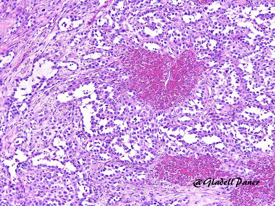
Striking example of pseudoangiosarcomatous (acantholytic) urothelial carcinoma. Pankeratin+, Gata3+, p63+, and vascular markers-. #GUPath

Striking example of pseudoangiosarcomatous (acantholytic) urothelial carcinoma. Pankeratin+, Gata3+, p63+, and vascular markers-. #GUPath
Ziad El-Zaatari (@ziad_zaatari) 's Twitter Profile Photo

🔬 "Fibrosis" ~ Trichrome stain of fibrosis in the renal pelvis of a patient with obstructive nephropathy ~ #PathArt 📷🎨

🔬 "Fibrosis" ~ Trichrome stain of fibrosis in the renal pelvis of a patient with obstructive nephropathy ~ #PathArt 📷🎨
Ankur Sangoi (@slusagar) 's Twitter Profile Photo

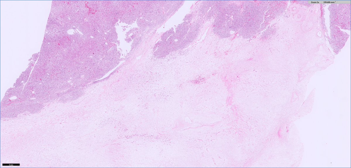
#GUpath #grosspath correlation: myxoid degeneration in nephrectomy of clear cell RCC 🚨👁️gross & histologic🔬mimic of tumor cell necrosis (an important poor prognostic factor for clear cell RCC)

#GUpath #grosspath correlation:

myxoid degeneration in nephrectomy of clear cell RCC

🚨👁️gross & histologic🔬mimic of tumor cell necrosis (an important poor prognostic factor for clear cell RCC)
Ziad El-Zaatari (@ziad_zaatari) 's Twitter Profile Photo

🔬📷 Diabetic Nodule ~ Takes over glomerulus as a vortex 🌪️ (PAS and Trichrome stains) ~ #RenalPath #Pathology #PathArt

🔬📷 Diabetic Nodule ~ Takes over glomerulus as a vortex 🌪️ (PAS and Trichrome stains) ~ #RenalPath #Pathology #PathArt
Arkana Laboratories (@arkanalabs) 's Twitter Profile Photo

This image (Jones silver stain) shows prominent glomerulitis in an allograft kidney biopsy, characterized in this case by increased mononuclear cells and segmental endothelial cell swelling within glomerular capillary loops. The patient is a young adult man who had received a

This image (Jones silver stain) shows prominent glomerulitis in an allograft kidney biopsy, characterized in this case by increased mononuclear cells and segmental endothelial cell swelling within glomerular capillary loops. The patient is a young adult man who had received a
Arkana Laboratories (@arkanalabs) 's Twitter Profile Photo

The light microscopic image depicts interstitial foam cells which, while non-specific and can be seen in longstanding proteinuria, are concerning for Alport Syndrome given the clinical scenario of hematuria and hearing loss. By electron microscopy, diagnostic findings would

Ankur Sangoi (@slusagar) 's Twitter Profile Photo

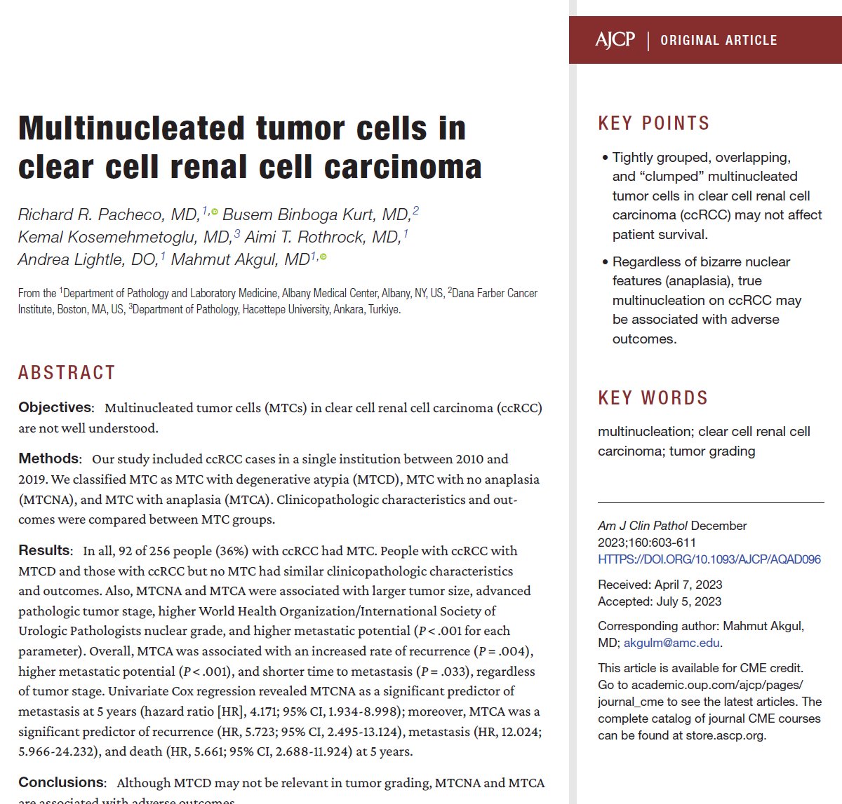
#GUpath grading of multinucleate tumor cells in RCC is a bit controversial - some apply WHO/ISUP and render grade 4, others grade 3/descriptive ✅some data but more needed! 🔬to me these are different than "syncytial-type" multinucleate giant cells which I consider bad/grade 4

#GUpath grading of multinucleate tumor cells in RCC is a bit controversial - some apply WHO/ISUP and render grade 4, others grade 3/descriptive

✅some data but more needed!
🔬to me these are different than "syncytial-type" multinucleate giant cells which I consider bad/grade 4
Grace Choung, MD 박영이 (@grace_choung) 's Twitter Profile Photo

Fibronectin glomerulopathy 🔸Massive fibronectin deposits 🔸AD mutation FN1 🔸MPGN-like pattern 🔸Silver-neg, PAS+, trichrome-red 🔸EM: Homogeneous granular + variable fibrils 🔸IF/IHC strongly + fibronectin 🔸renal-limited (1/2) #renalpath #PathTwitter

Fibronectin glomerulopathy
🔸Massive fibronectin deposits
🔸AD mutation FN1
🔸MPGN-like pattern
🔸Silver-neg, PAS+, trichrome-red
🔸EM: Homogeneous granular + variable fibrils
🔸IF/IHC strongly + fibronectin
🔸renal-limited
(1/2)
#renalpath #PathTwitter
Ziad El-Zaatari (@ziad_zaatari) 's Twitter Profile Photo

🔬📷 Beautiful section of the Epididymis ~ Can you identify the key areas seen in the image? ☑️ Efferent Ductules ☑️ Head ⭐️ Bonus! A small Appendix Epididymis #GUpath #Histology #Pathology #Testis #PathologyBasics

🔬📷 Beautiful section of the Epididymis ~ Can you identify the key areas seen in the image?  
   ☑️ Efferent Ductules
   ☑️ Head
   ⭐️ Bonus! A small Appendix Epididymis
#GUpath #Histology #Pathology #Testis #PathologyBasics
Arkana Laboratories (@arkanalabs) 's Twitter Profile Photo

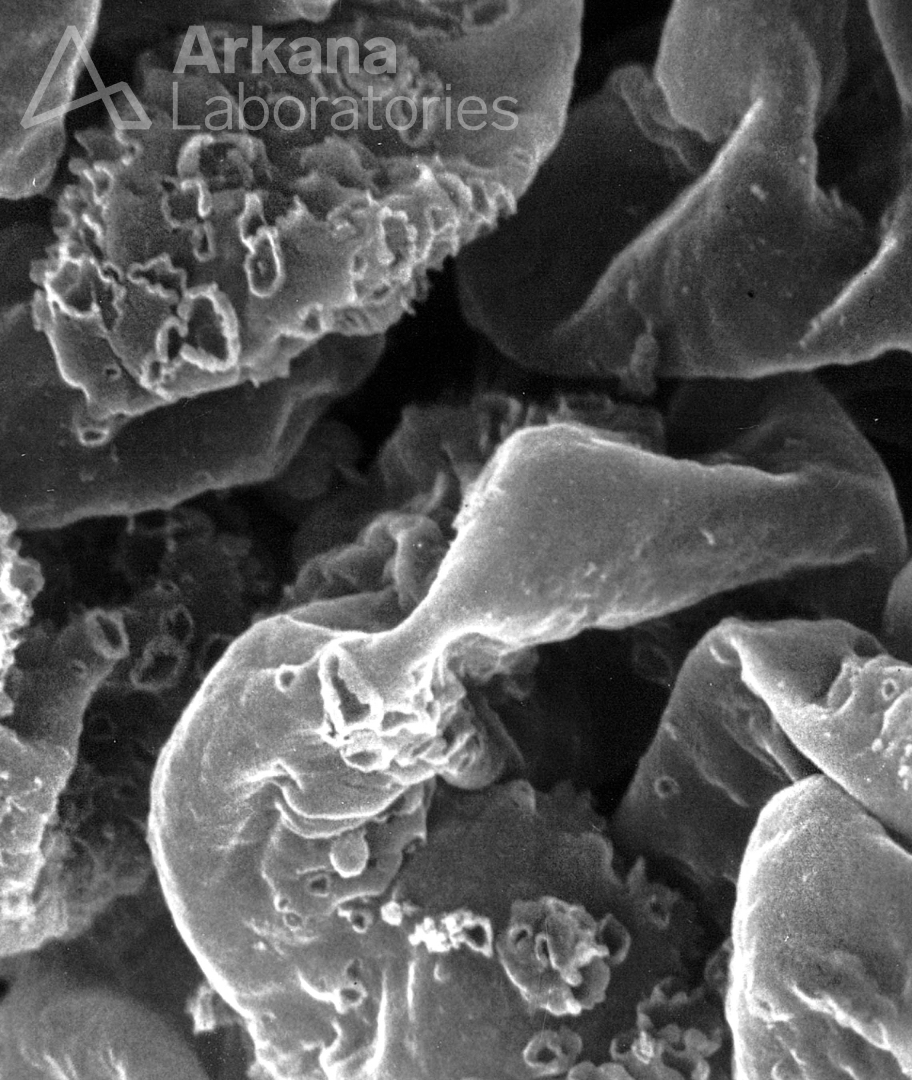
Today's #eyeSCANdy is a SEM of acellular GBMs from segmental membranous illustrating the variability of size and shape of the GBM formations.  Photo courtesy of Dr. Stephen Bonsib. #renal #pathology #kidneypath

Today's #eyeSCANdy is a SEM of acellular GBMs from segmental membranous illustrating the variability of size and shape of the GBM formations. 

Photo courtesy of Dr. Stephen Bonsib. #renal #pathology #kidneypath
Ankur Sangoi (@slusagar) 's Twitter Profile Photo

for the #pathresidents: #GUpath radical prostatectomy for bx-proven 3+4 prostate cancer; patient also has h/o bladder cancer

for the #pathresidents:

#GUpath radical prostatectomy for bx-proven 3+4 prostate cancer; patient also has h/o bladder cancer
Human Pathology (@human_pathology) 's Twitter Profile Photo

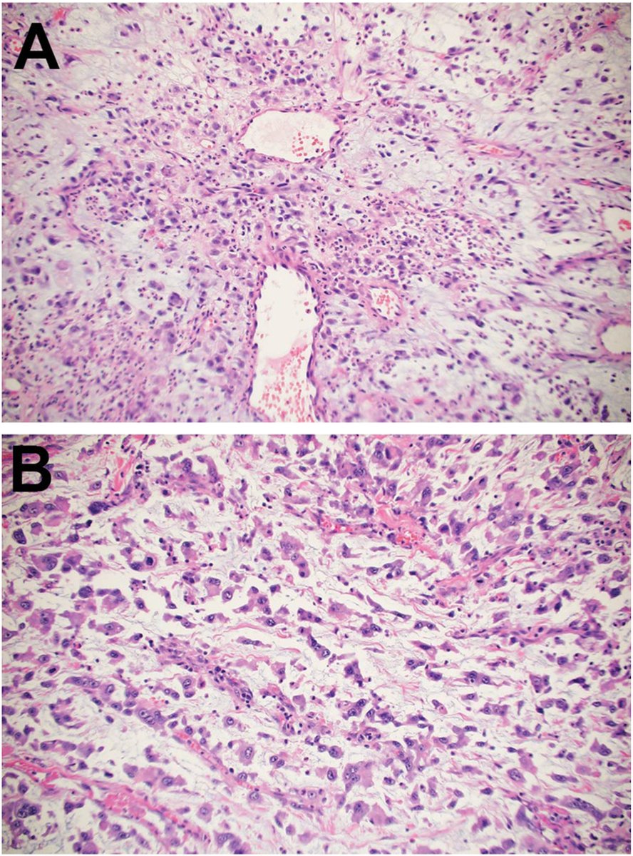
New in #HumPathol: Invasive urothelial carcinoma with chordoid and myxoid features shows increased RAS/RAF pathway alterations. sciencedirect.com/science/articl… #pathology #PathTwitter #PathX #GUpath

New in #HumPathol: Invasive urothelial carcinoma with chordoid and myxoid features shows increased RAS/RAF pathway alterations. sciencedirect.com/science/articl… #pathology #PathTwitter #PathX #GUpath
Jonathan Zuckerman MD PhD (@jzrenalpath) 's Twitter Profile Photo

One of the more subtle amyloid cases I have seen. Just a few deposits in 2 gloms by LM. Just enough congo red staining. Glad to catch it in the EM as well. #renalpath #nephrology #pathtwitter

One of the more subtle amyloid cases I have seen. Just a few deposits in 2 gloms by LM. Just enough congo red staining. Glad to catch it in the EM as well.   #renalpath #nephrology #pathtwitter
Ziad El-Zaatari (@ziad_zaatari) 's Twitter Profile Photo

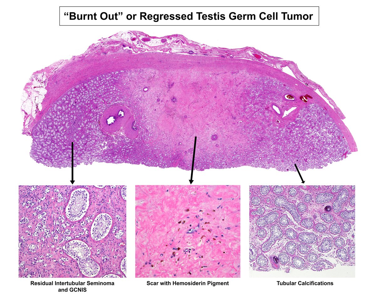
🔬Case of a "Burnt Out" or Regressed Germ Cell Tumor: ➡️ Fibrous scar (middle) with hemosiderin pigment ➡️ Some residual seminoma and GCNIS (left) ➡️ Intratubular calcifications (seen focally on right) #GUpath #Pathology #Testis #TesticularCancer

🔬Case of a "Burnt Out" or Regressed Germ Cell Tumor: 
   ➡️ Fibrous scar (middle) with hemosiderin pigment
   ➡️ Some residual seminoma and GCNIS (left)
   ➡️ Intratubular calcifications (seen focally on right)
#GUpath #Pathology #Testis #TesticularCancer
Arkana Laboratories (@arkanalabs) 's Twitter Profile Photo

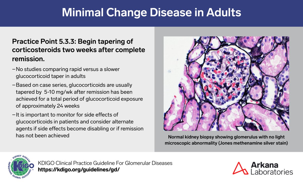
A sample from the pathology teaching slide set on Minimal Change Disease in Adults, jointly prepared by Arkana Laboratories and KDIGO. KDIGO.org/guidelines/gd/ #KDIGO #pathology #renal #renalpath #pathx #pathtwitter

A sample from the pathology teaching slide set on Minimal Change Disease in Adults, jointly prepared by Arkana Laboratories and <a href="/goKDIGO/">KDIGO</a>. KDIGO.org/guidelines/gd/

#KDIGO #pathology #renal #renalpath #pathx #pathtwitter
Ankur Sangoi (@slusagar) 's Twitter Profile Photo

#GUpath radical nephrectomy for RCC grossly encroaching renal pelvis: is top vs bottom row pelvicalyceal invasion (PCI; which is latest pT3a addition to AJCC staging)? and what constitutes PCI? in👇🏼interobserver study, Sean R Williamson MD & I explore this, offering our $0.02

#GUpath radical nephrectomy for RCC grossly encroaching renal pelvis:

is top vs bottom row pelvicalyceal invasion (PCI; which is latest pT3a addition to AJCC staging)?

and what constitutes PCI?

in👇🏼interobserver study, <a href="/Williamson_SR/">Sean R Williamson MD</a> &amp; I explore this, offering our $0.02
Ankur Sangoi (@slusagar) 's Twitter Profile Photo

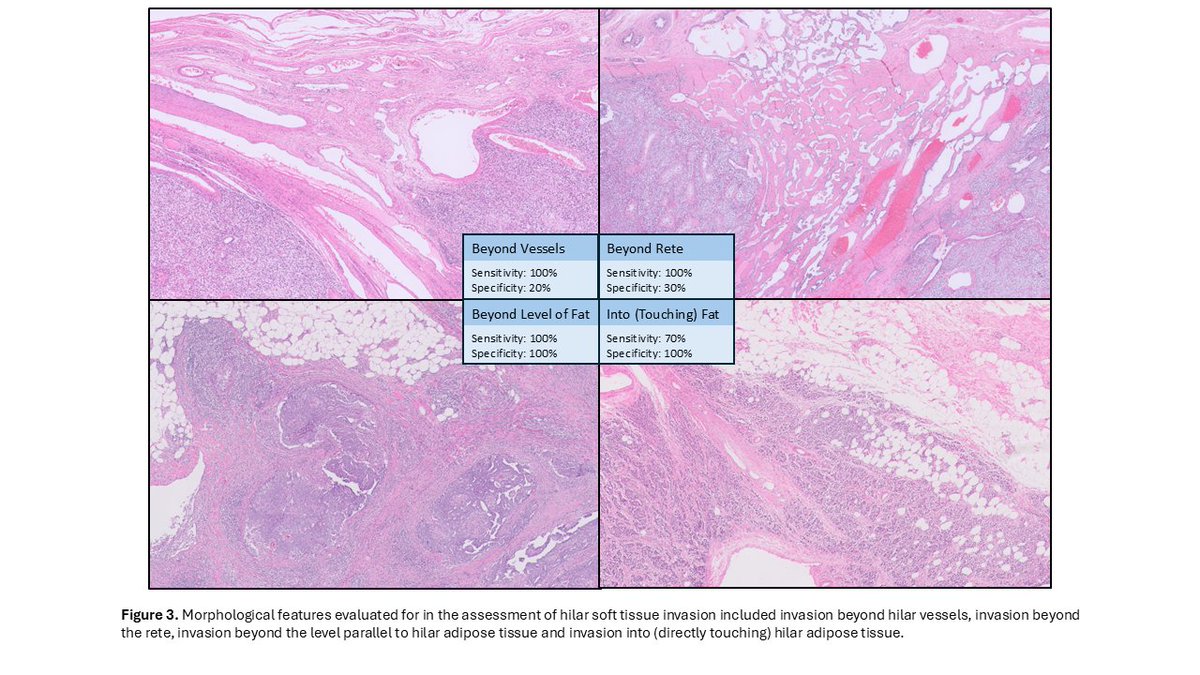
#GUpath orchiectomy for germ cell tumor grossly encroaching hilum: should top vs bottom row be considered "hilar soft tissue invasion" (HSTI)? HSTI=pT2, so critical staging parameter! how should HSTI be defined? in👇🏽interobserver study, we investigate this and offer some💡

#GUpath orchiectomy for germ cell tumor grossly encroaching hilum:

should top vs bottom row be considered "hilar soft tissue invasion" (HSTI)?  HSTI=pT2, so critical staging parameter!

how should HSTI be defined?

in👇🏽interobserver study, we investigate this and offer some💡
Ziad El-Zaatari (@ziad_zaatari) 's Twitter Profile Photo

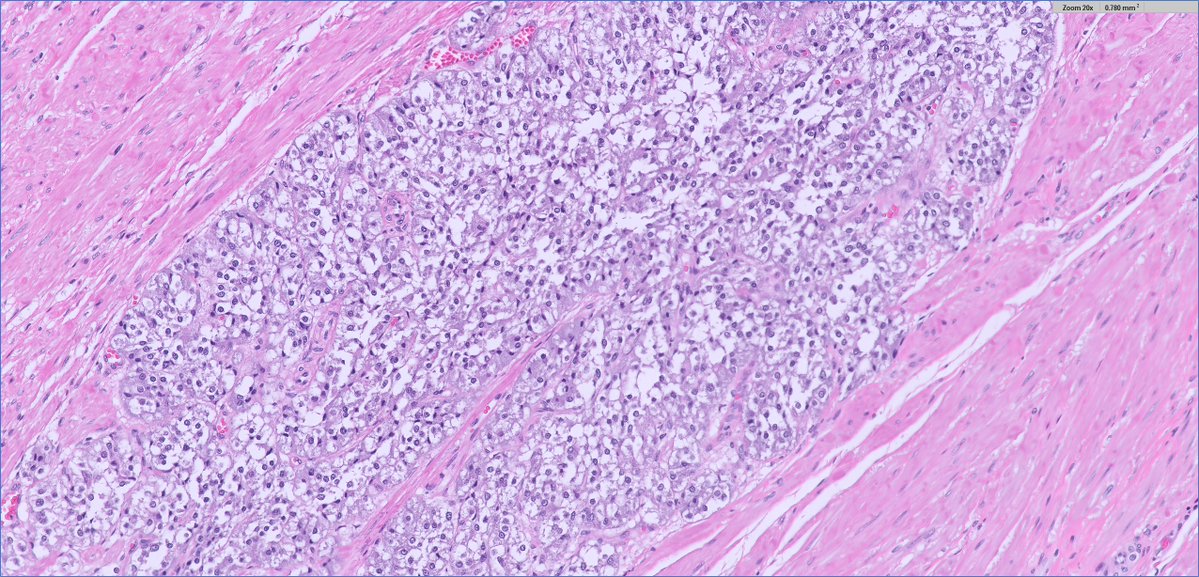
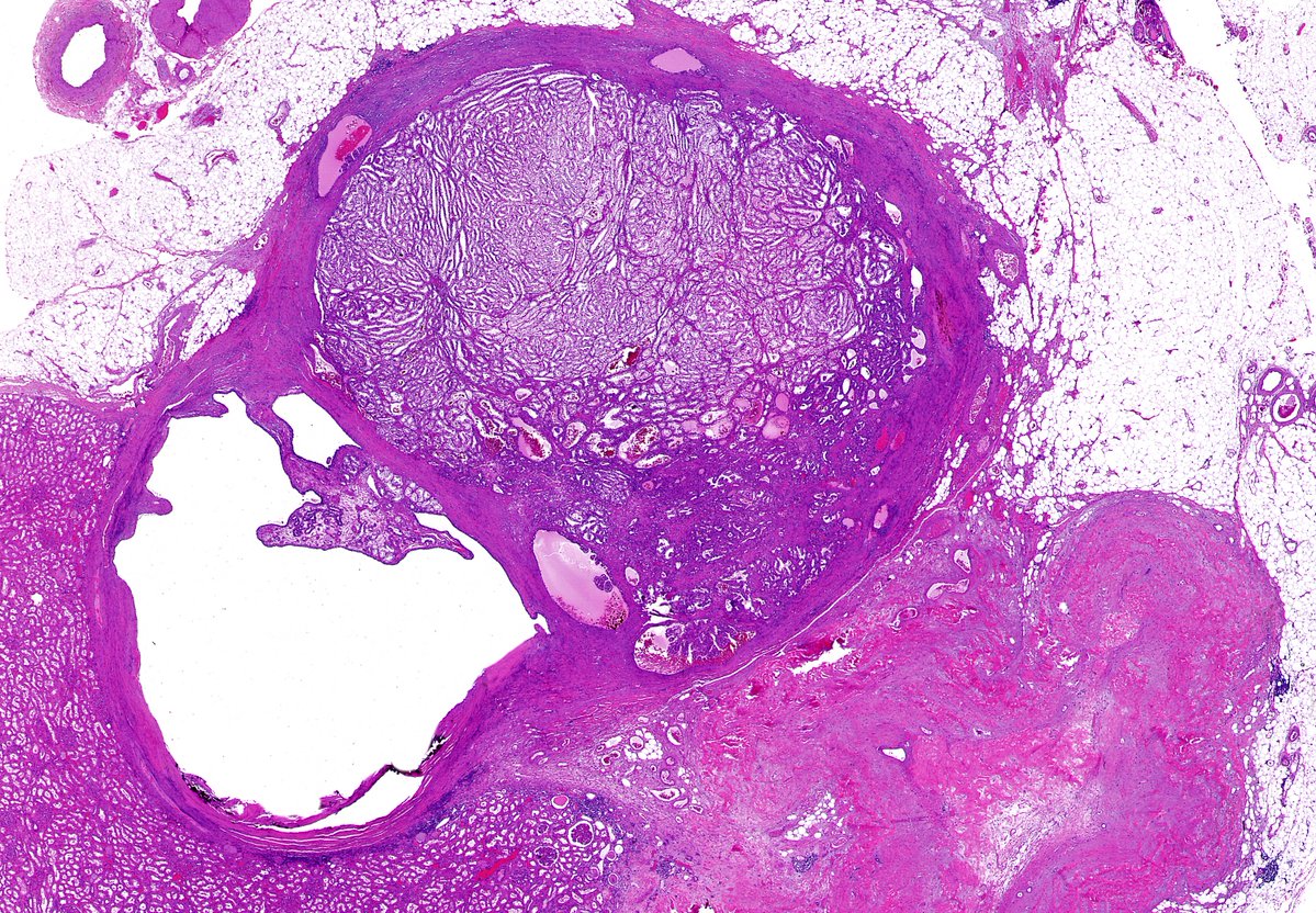
🔬 Can you identify this renal tumor in this low power view? Or is it #TooLowToDiagnose? #GUpath #Pathology #Kidney #KidneyTumors

🔬 Can you identify this renal tumor in this low power view? Or is it #TooLowToDiagnose? #GUpath #Pathology #Kidney #KidneyTumors