Vascular Surgery Associates (@vascsurgla) 's Twitter Profile
Vascular Surgery Associates

@vascsurgla

Willis Wagner, MD
David Cossman, MD
Rajeev Rao, MD
Allan Tulloch, MD
Ryan Haqq, MD
Rameen Moridzadeh, MD

ID: 1148649762721189888

linkhttp://www.vascularsurg.com calendar_today09-07-2019 17:46:59

18 Tweet

164 Followers

63 Following

Vascular Surgery Associates (@vascsurgla) 's Twitter Profile Photo

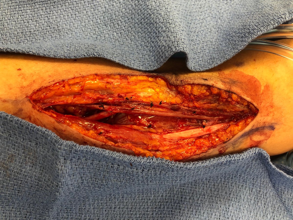
Spontaneous proximal ulnar artery aneurysm in a young patient treated with interposition vein graft. No evidence of connective tissue disease. #vascularsurgery #aneurysm #bypass

Spontaneous proximal ulnar artery aneurysm in a young patient treated with interposition vein graft. No evidence of connective tissue disease. #vascularsurgery #aneurysm #bypass
Vascular Surgery Associates (@vascsurgla) 's Twitter Profile Photo

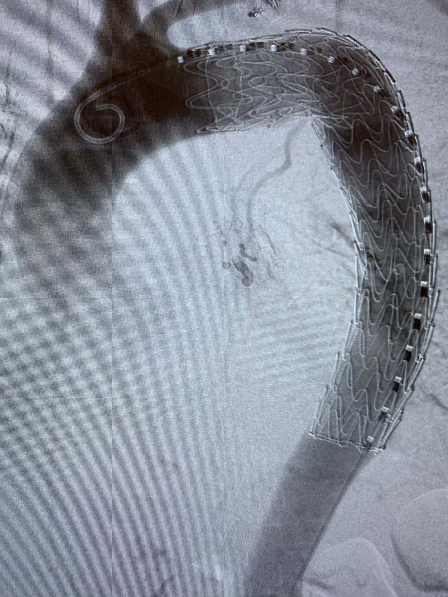
Bilateral carotid subclavian transposition and TEVAR for aberrant right subclavian artery with kommerell’s diverticulum. This patient also had a common origin for the innominate and L CCA. #vascularsurgery #TEVAR

Bilateral carotid subclavian transposition and TEVAR for aberrant right subclavian artery with kommerell’s diverticulum. This patient also had a common origin for the innominate and L CCA. #vascularsurgery #TEVAR
Vascular Surgery Associates (@vascsurgla) 's Twitter Profile Photo

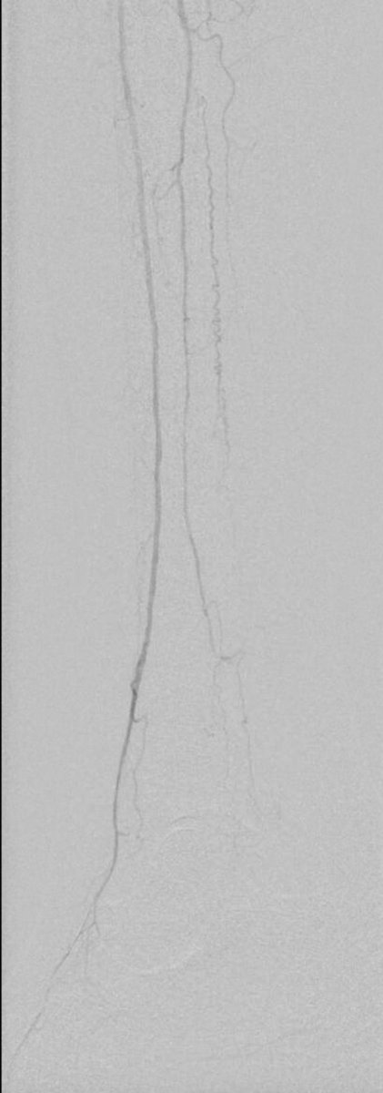
69 year old woman with a history of heart transplant with right great toe gangrene and segmental occlusion in the anterior tibial artery and dorsalis pedis - before and after treatment.

69 year old woman with a history of heart transplant with right great toe gangrene and segmental occlusion in the anterior tibial artery and dorsalis pedis - before and after treatment.
Vascular Surgery Associates (@vascsurgla) 's Twitter Profile Photo

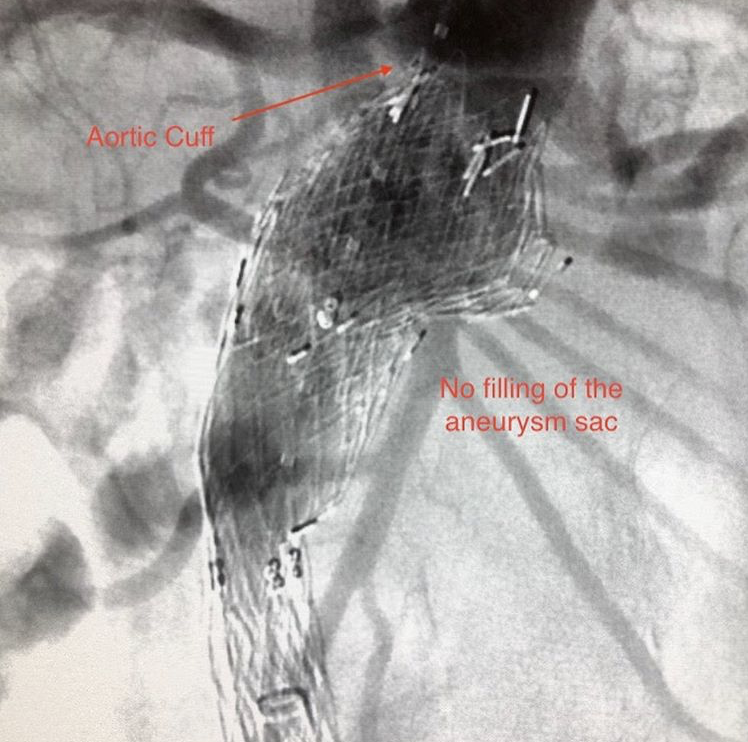
This patient presented with a symptomatic type 1a endoleak after EVAR. The patient was treated with aortic cuffs to bridge to the renal arteries. The second picture shows the absence of contrast in the aneurysm sac and adequate seal at the renal arteries. #vascularsurgery #EVAR

This patient presented with a symptomatic type 1a endoleak after EVAR. The patient was treated with aortic cuffs to bridge to the renal arteries. The second picture shows the absence of contrast in the aneurysm sac and adequate seal at the renal arteries. #vascularsurgery #EVAR
Vascular Surgery Associates (@vascsurgla) 's Twitter Profile Photo

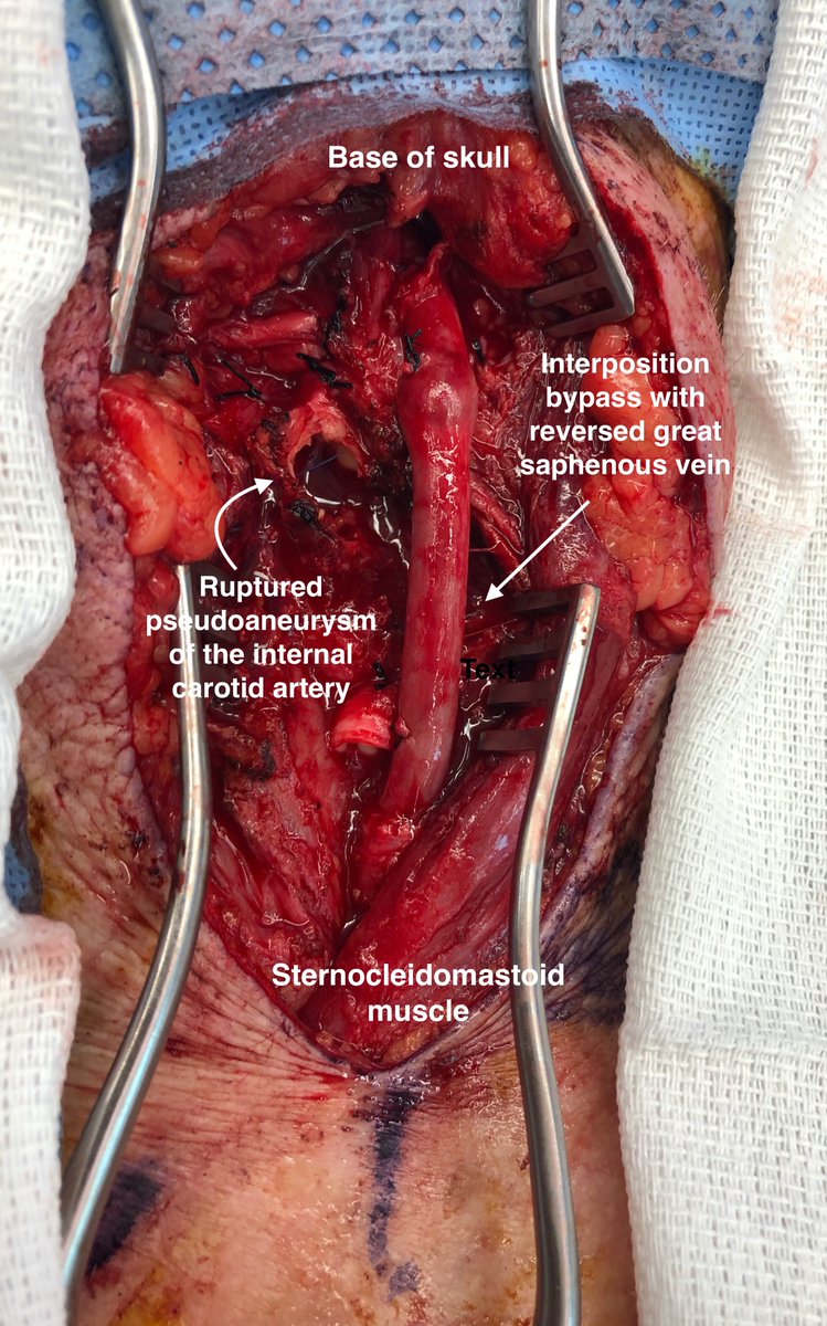
This patient presented to the hospital with bacteremia and enlarging neck mass. The patient was found to have a contained, ruptured pseudoaneurysm of the ICA. This was repaired with interposition bypass with saphenous vein and ligation of the ECA. #vascularsurgery #pseudoaneurysm

This patient presented to the hospital with bacteremia and enlarging neck mass. The patient was found to have a contained, ruptured pseudoaneurysm of the ICA. This was repaired with interposition bypass with saphenous vein and ligation of the ECA. #vascularsurgery #pseudoaneurysm
Vascular Surgery Associates (@vascsurgla) 's Twitter Profile Photo

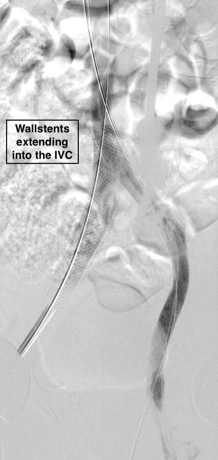
This patient had an #IVCF placed >10 years ago and presented with occluded left iliac #veins and large pelvic collaterals and leg edema. The patient underwent filter retrieval and iliocaval reconstruction with wallstents. The pelvic collaterals/leg edema have resolved! #vascular

This patient had an #IVCF placed >10 years ago and presented with occluded left iliac #veins and large pelvic collaterals and leg edema. The patient underwent filter retrieval and iliocaval reconstruction with wallstents. The pelvic collaterals/leg edema have resolved! #vascular
Vascular Surgery Associates (@vascsurgla) 's Twitter Profile Photo

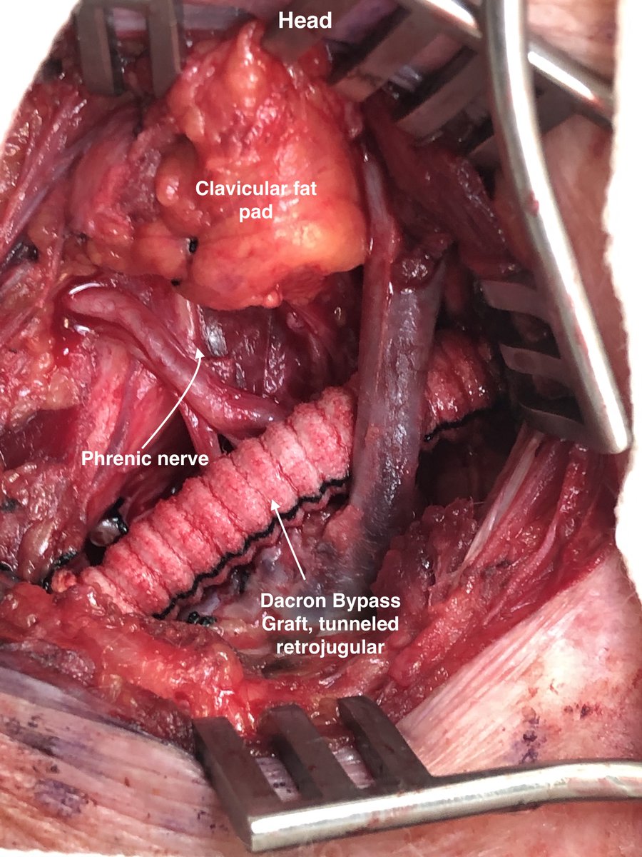
This patient presented with progressive dry necrosis to the fingertips. Angiogram showed severe proximal stenosis. Given the proximity to the vertebral artery, a stent was not placed. This patient was treated with carotid-subclavian bypass with ligation of SCA. #vascularsurgery

This patient presented with progressive dry necrosis to the fingertips. Angiogram showed severe proximal stenosis. Given the proximity to the vertebral artery, a stent was not placed. This patient was treated with carotid-subclavian bypass with ligation of SCA. #vascularsurgery
Vascular Surgery Associates (@vascsurgla) 's Twitter Profile Photo

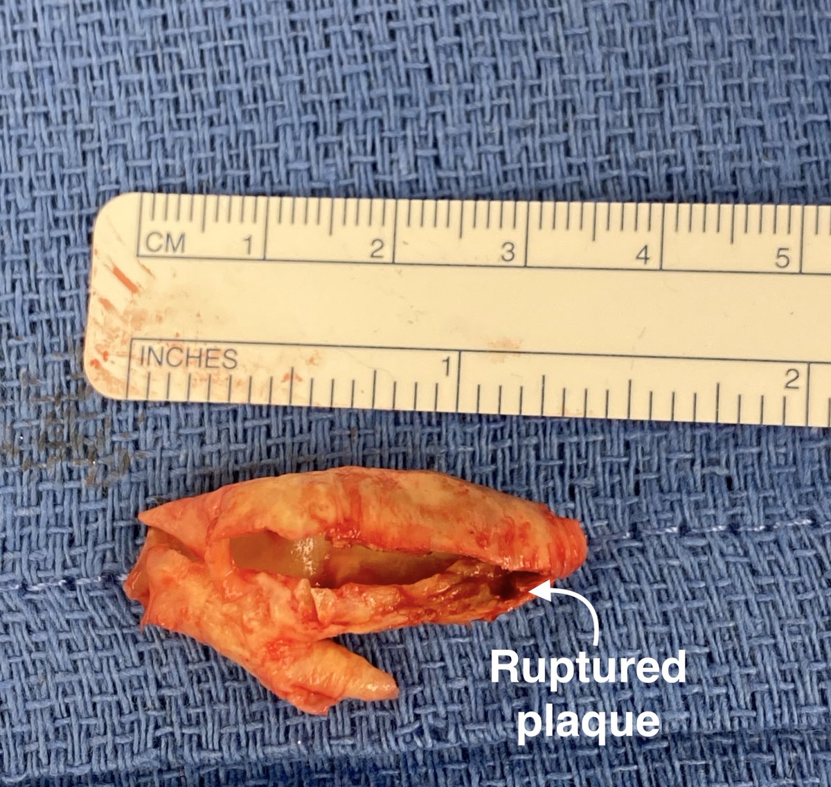
This patient presented with a symptomatic #carotid stenosis. CTA showed a string sign of the ICA. The patient underwent #carotidendarterectomy which revealed a ruptured plaque of the ICA. The artery was patched and the patient did well postoperatively.

This patient presented with a symptomatic #carotid stenosis.  CTA showed a string sign of the ICA. The patient underwent #carotidendarterectomy which revealed a ruptured plaque of the ICA. The artery was patched and the patient did well postoperatively.
Rameen S. Moridzadeh, MD (@moridzadehmd) 's Twitter Profile Photo

This patient presented with sudden onset of left leg swelling. CT venogram demonstrated iliofemoral DVT with #thrombosis down to the femoral vein. Treated single session using JETi thrombectomy followed by IVUS and iliac vein stenting. #MedEd Vascular Surgery Associates
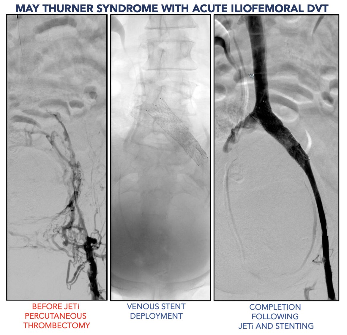
This patient presented with sudden onset of left leg swelling. CT venogram demonstrated iliofemoral DVT with #thrombosis down to the femoral vein. Treated single session using JETi thrombectomy followed by IVUS and iliac vein stenting. #MedEd <a href="/VascSurgLA/">Vascular Surgery Associates</a>
DanielMilesMD (@danielmilesmd) 's Twitter Profile Photo

Patient with chronic mesenteric ischemia with unsuccessful endovascular attempt treated with aorta to SMA bypass (Dacron). Replaced common hepatic from SMA! Vascular Surgery Associates Cedars-Sinai Division of Vascular Surgery Department of Surgery at Cedars-Sinai #vascularsurgery #SoMe4Surgery #MedEd
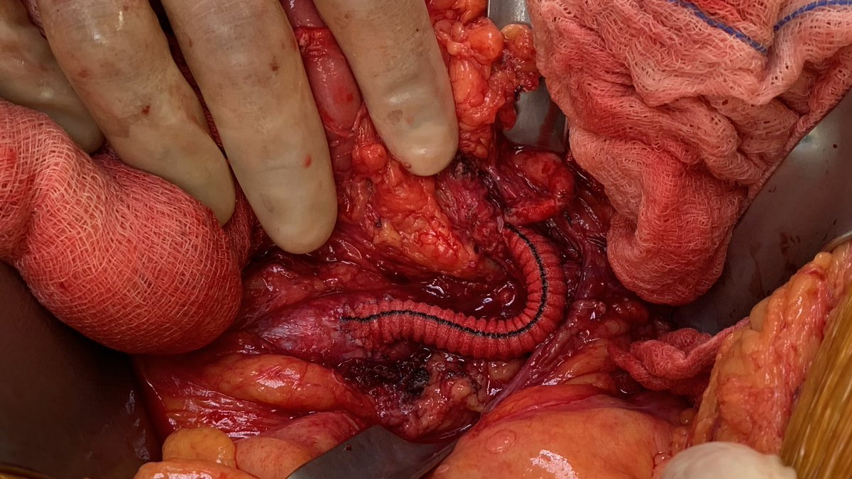
Patient with chronic mesenteric ischemia with unsuccessful endovascular attempt treated with aorta to SMA bypass (Dacron). Replaced common hepatic from SMA! <a href="/VascSurgLA/">Vascular Surgery Associates</a> <a href="/VascularSurgCS/">Cedars-Sinai Division of Vascular Surgery</a> <a href="/DeptSurgeryCS/">Department of Surgery at Cedars-Sinai</a> #vascularsurgery #SoMe4Surgery #MedEd