Ash Aryal (@dallasyeti) 's Twitter Profile
Ash Aryal

@dallasyeti

Confocal & Super Resolution Applications Specialist, Biomedical Engineer, Zeiss Microscopy, Views are my own and not of my employer

ID: 714245038

calendar_today14-10-2013 10:31:04

151 Tweet

154 Followers

1,1K Following

Hyser Lab (@hyserlab) 's Twitter Profile Photo

#WonderfulWednesday on the #Zeiss #Airyscan2 watching #calciumsignaling ⚡️-amazed by the diversity of the types of Ca2+ signals in #rotavirus infected cells. Small and large Ca2+ puffs & intracellular Ca2+ waves…others? (*GCaMP6s Ca2+ sensor, ~3Hz imaging for 10 min)

Institute for Chemical Imaging of Living Systems (@cilsinstitute) 's Twitter Profile Photo

Image of the Week: Brain microvascular network - brain endothelial cells (red), pericytes (purple), and astrocytes (green) - imaged on the ZEISS Microscopy LSM 800. #FluorescenceFriday #Imaging #Microscopy Credit: Max Winkelman/Dai Lab


Image of the Week: Brain microvascular network - brain endothelial cells (red), pericytes (purple), and astrocytes (green) - imaged on the <a href="/zeiss_micro/">ZEISS Microscopy</a> LSM 800.
#FluorescenceFriday #Imaging #Microscopy

Credit: Max Winkelman/Dai Lab
ZEISS Microscopy (@zeiss_micro) 's Twitter Profile Photo

Introducing a new confocal experience. Join us for a special event stream on December 8: zeiss.ly/tw-new-confoca… #confocal #microscopy #airyscan #imaging #NewConfocalExperience #stream

Nat Prunet 🌸🔬🏳️‍🌈 (@nat_prunet) 's Twitter Profile Photo

#ant imaged on ZEISS Microscopy LSM880 #confocal #microscope. #Autofluorescence. Those eyes are striking! #microscopy #bioart #sciart #microscopyart #microscopicworld #closeup #biology #insects
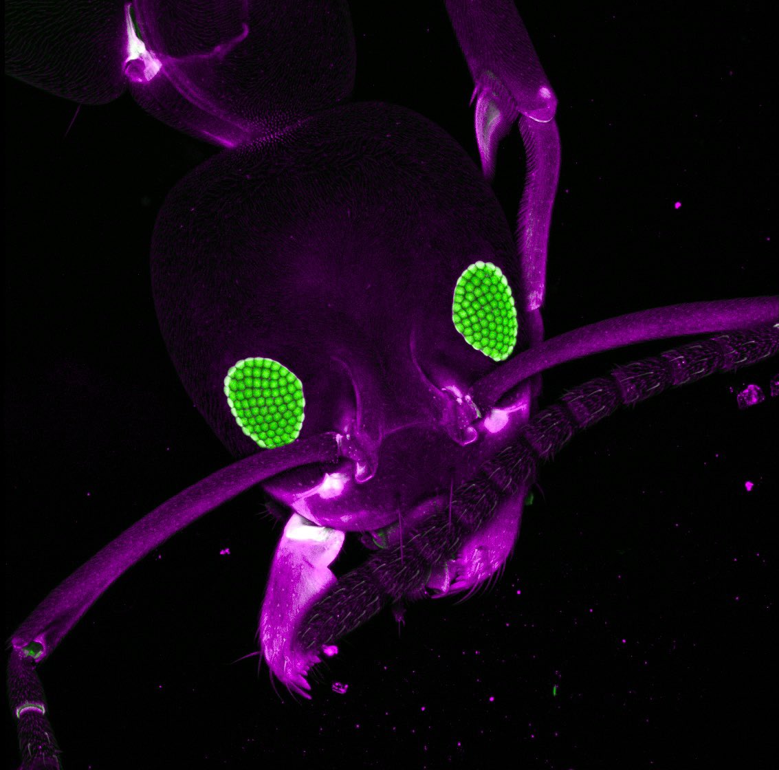
#ant imaged on <a href="/zeiss_micro/">ZEISS Microscopy</a> LSM880 #confocal #microscope. #Autofluorescence. Those eyes are striking!

#microscopy #bioart #sciart #microscopyart #microscopicworld #closeup #biology #insects
Chih-Wei Logan Hsu (@cwloganhsu) 's Twitter Profile Photo

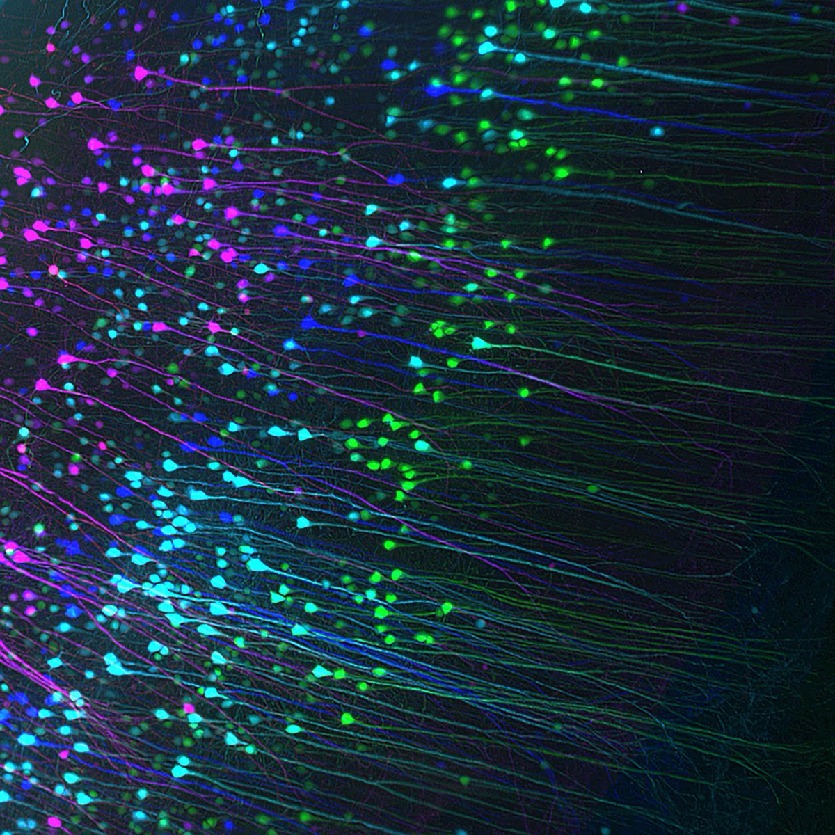
Color-coded depth projection of Thy1-EGFP+ cells from EZ Clear processed adult mouse brain imaged on Zeiss Lightsheet Z.1! #tissueclearing #3Dimaging #Zeiss #lightsheet

Color-coded depth projection of Thy1-EGFP+ cells from EZ Clear processed adult mouse brain imaged on Zeiss Lightsheet Z.1!
#tissueclearing #3Dimaging #Zeiss #lightsheet
Akaash Kumar (@_akaash_kumar) 's Twitter Profile Photo

My contribution to #fluorescencefriday -fly notum imaged on the ZEISS Microscopy Lattice Light Sheet. Sample prepared by Alice Bittleston in the @DeriveryLab and napari used for processing/animation.

ZEISS Microscopy (@zeiss_micro) 's Twitter Profile Photo

What do all the life science images in this thread (1/11) have in common? They have been acquired by users in our incredible social media community, with ZEISS microscopes, throughout the year 2021! 🙏🔬 Read the full thread to learn more about all nine most popular images 👇

What do all the life science images in this thread (1/11) have in common? They have been acquired by users in our incredible social media community, with ZEISS microscopes, throughout the year 2021! 🙏🔬

Read the full thread to learn more about all nine most popular images 👇
Sara Di Rienzi (@saradirienzi) 's Twitter Profile Photo

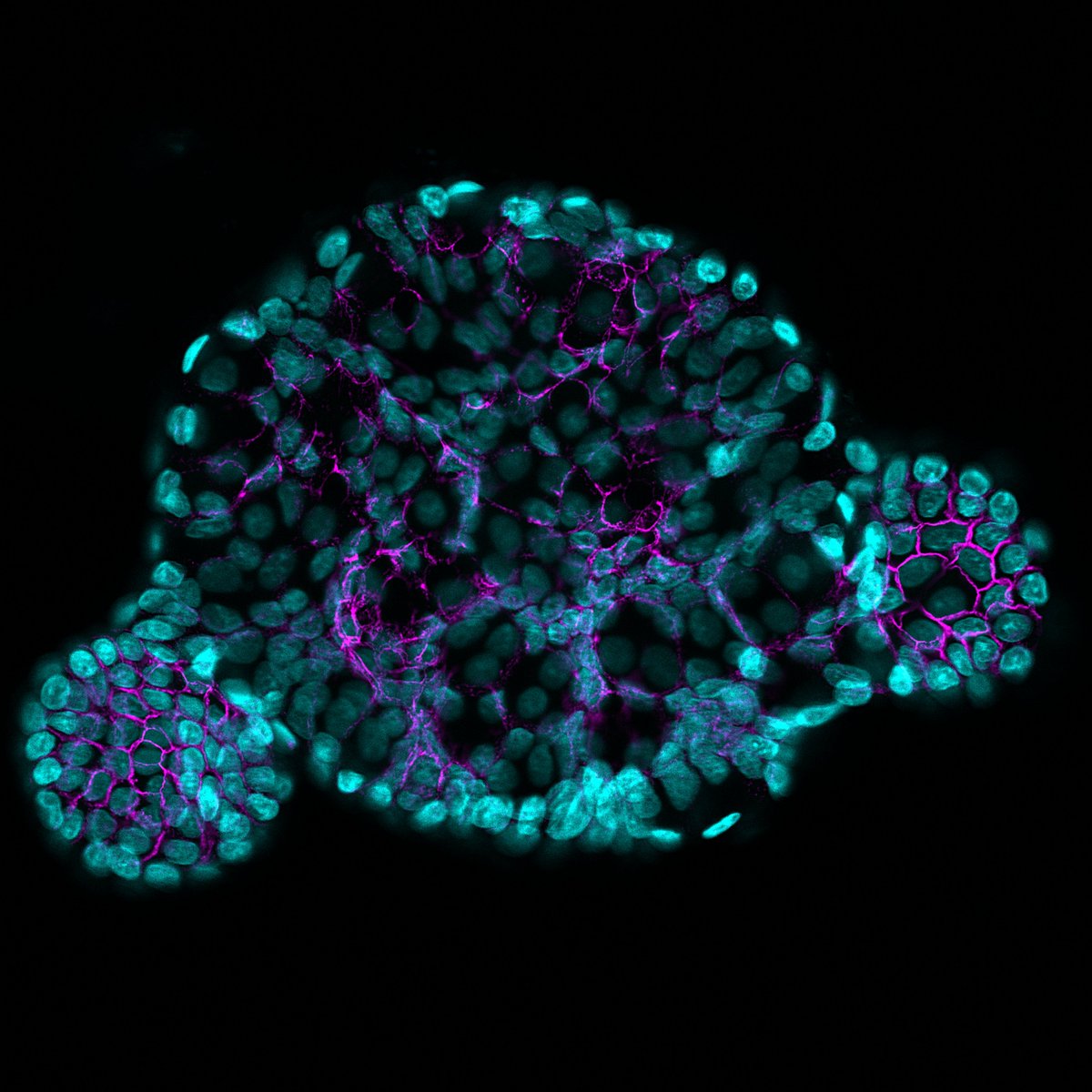
Baby enteroid! 🥰 Infant derived jejunal enteroid stained with DAPI (cyan) and E-cadherin in purple. Imaged with the glorious AiryScan. #FluorescenceFriday

Baby enteroid! 🥰 Infant derived jejunal enteroid stained with DAPI (cyan) and E-cadherin in purple. Imaged with the glorious AiryScan. #FluorescenceFriday
Sébastien Mailfert (@mailfertseb) 's Twitter Profile Photo

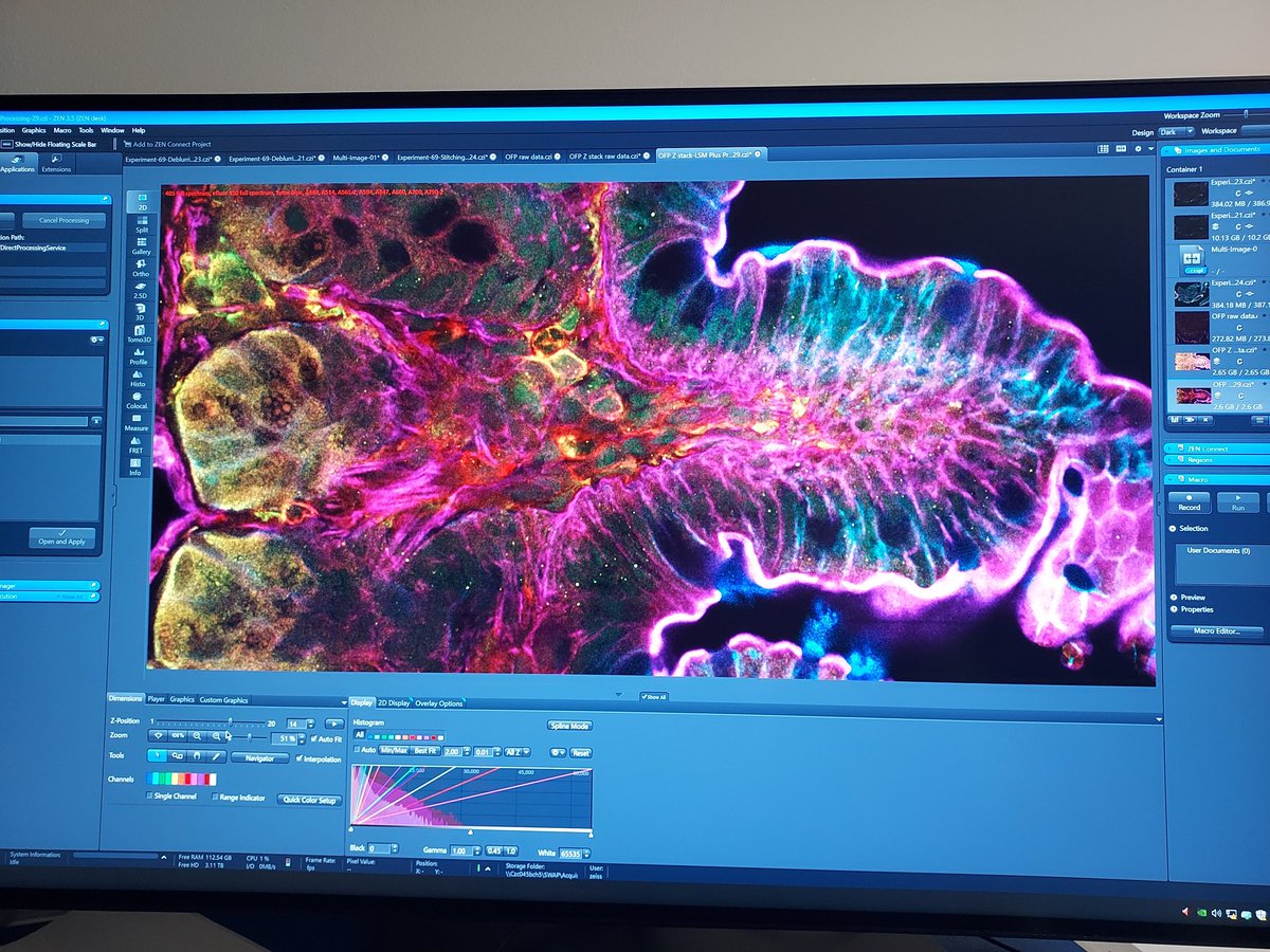
11 colors acquired during the on-site demo of the brand new Zeiss LSM980 with spectral detection and infrared detectors with LSMPlus... just AMAZING! Sample from Hugues Lelouard, CIML CNRS 🌍 Inserm @univamu ZEISS Microscopy

11 colors acquired during the on-site demo of the brand new Zeiss LSM980 with spectral detection and infrared detectors with LSMPlus... just AMAZING! Sample from Hugues Lelouard, <a href="/CIML_Immunology/">CIML</a> <a href="/CNRS/">CNRS 🌍</a> <a href="/Inserm/">Inserm</a> @univamu <a href="/zeiss_micro/">ZEISS Microscopy</a>
Bret Judson (@judsonlab) 's Twitter Profile Photo

For your #FluorescenceFriday consideration here is a ZEISS Microscopy Airyscan image of microtubules and a nucleus in Drosophila muscle. Never get tired of imaging these. With Eric Folker and Boston College
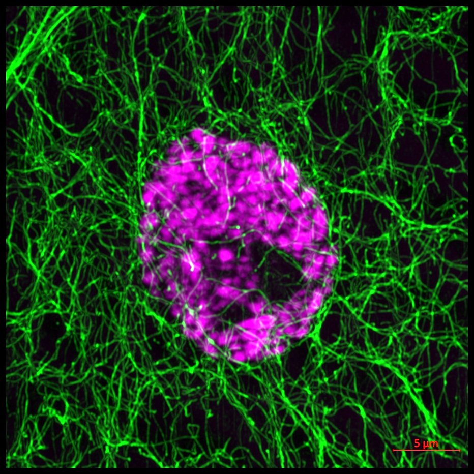
For your #FluorescenceFriday consideration here is a <a href="/zeiss_micro/">ZEISS Microscopy</a> Airyscan image of microtubules and a nucleus in Drosophila muscle.  Never get tired of imaging these.  With <a href="/folker_lab/">Eric Folker</a> and <a href="/BostonCollege/">Boston College</a>
Katie Smith (@k_e_smith86) 's Twitter Profile Photo

So nice when you manage to find your dye-filled cell after immunolabelling! Here's a glial cell (orange) reaching out to several different axons (blue). Airyscan imaging ZEISS Microscopy UCL Ear Institute #FluorescenceFriday
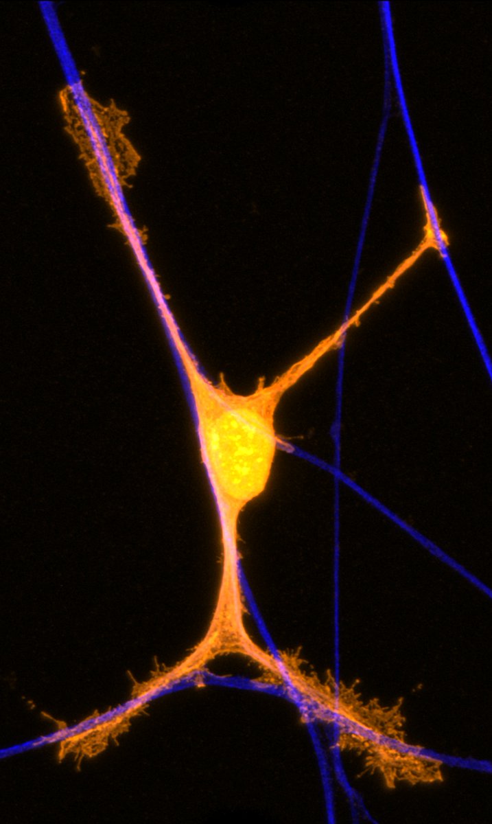
So nice when you manage to find your dye-filled cell after immunolabelling! Here's a glial cell (orange) reaching out to several different axons (blue). Airyscan imaging <a href="/zeiss_micro/">ZEISS Microscopy</a> <a href="/UCLEarInstitute/">UCL Ear Institute</a>  #FluorescenceFriday
Chih-Wei Logan Hsu (@cwloganhsu) 's Twitter Profile Photo

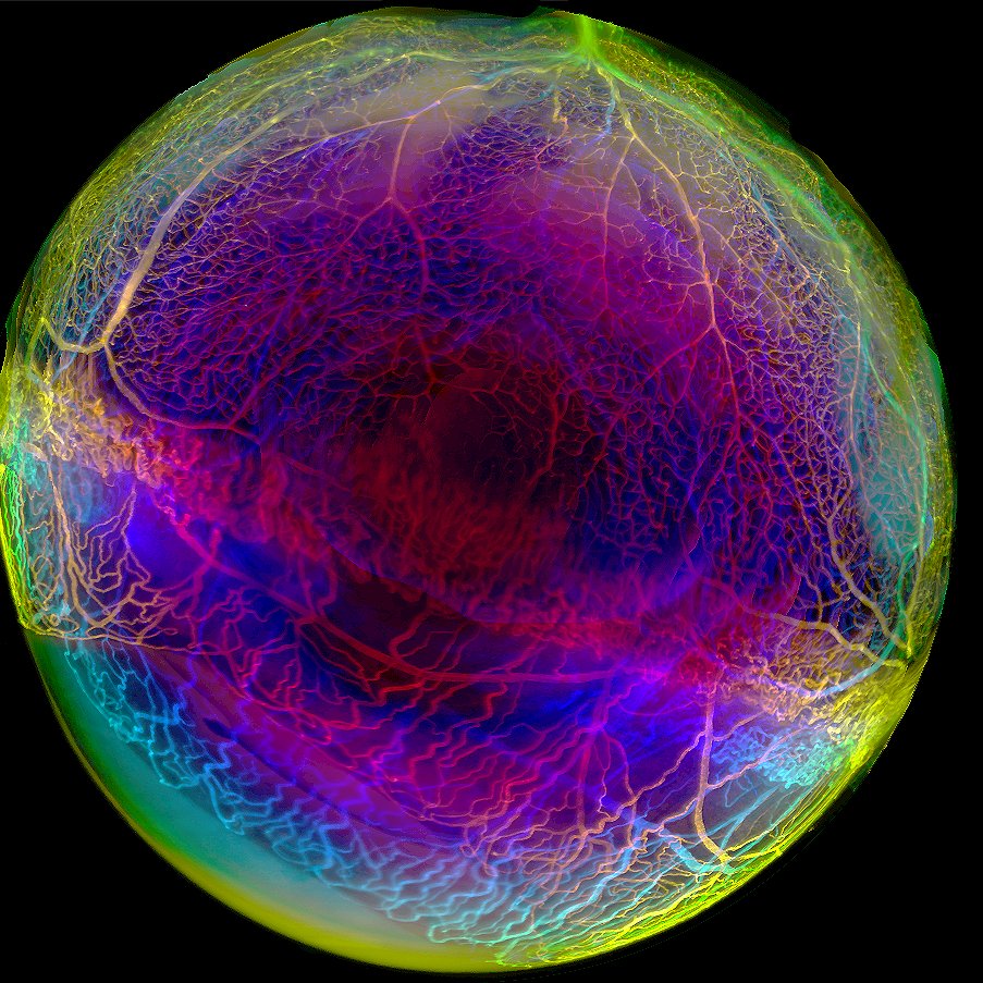
Another "eye" candy captured on lightsheet. Cleared mouse whole eye processed by #EZclear and imaged on #Zeiss Lightsheet Z.1. Look at that retinal vasculature 😍! Happy #FluorescenceFriday!

Another "eye" candy captured on lightsheet. Cleared mouse whole eye processed by #EZclear and imaged on #Zeiss Lightsheet Z.1. Look at that retinal vasculature 😍! Happy #FluorescenceFriday!
Chih-Wei Logan Hsu (@cwloganhsu) 's Twitter Profile Photo

Very impressed with the data from the test run on ZEISS Microscopy Xradia Context microCT by Dr. Mansoureh Norouzi Rad at ZMCC BA! This iodine contrasted E15.5 mouse embryo data was imaged at 4.3 um/voxel with rendering & animation in Imaris 3D/4D Imaging. So much detail to look at!

Chih-Wei Logan Hsu (@cwloganhsu) 's Twitter Profile Photo

CD31+ embryonic vascular system in a E12.5 mouse embryo. Sample was immunostained and processed with #EZClear by Dr. Nanbing Jade Li-Villarreal Jade Li BCM Department of Integrative Physiology and imaged on ZEISS Microscopy Lightsheet Z.1 by #OiVM BCMHouston. Happy #FluorescenceFriday 😎!

Collene Jeter (@collene_jeter) 's Twitter Profile Photo

Wrapping up an exciting 3 week demo of the Zeiss Lattice Lightsheet 7 here at MDACC Advanced Microscopy Core. Shout out to Ash Aryal (Ash Aryal) and David Akalal for outstanding support. Image: focal adhesion (Paxillin-mCherry) live murine lung cancer #microscopy #zeiss_micro