Diego Lemos (@dflemosmd) 's Twitter Profile
Diego Lemos

@dflemosmd

Division Chief of Musculoskeletal Imaging at the University of Vermont Medical Center #mskrad

ID: 3062170906

calendar_today25-02-2015 14:26:23

7,7K Tweet

4,4K Followers

1,1K Following

James Noake (@drjn_sportsmed) 's Twitter Profile Photo

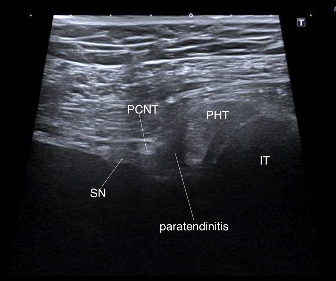
Consent ✅ Training for marathon - ramp in volume / distance Initially inferior gluteal pain Now evolved into more diffuse, neuropathic radiating 'ache' into posterior thigh to knee, but not below See pain map Worse with sitting, long leg stretch, inclines POCUS - proximal

Consent ✅

Training for marathon - ramp in volume / distance

Initially inferior gluteal pain

Now evolved into more diffuse, neuropathic radiating 'ache' into posterior thigh to knee, but not below

See pain map

Worse with sitting, long leg stretch, inclines 

POCUS - proximal
MSK Teaching Room (@mskteachingroom) 's Twitter Profile Photo

24 y/o guy 🤵‍♂️ with pain in the left groin 😣 link to case ⬇️ pacsbin.com/c/ZJAEz-E32o what is your differential? Weekend vibes! Enjoy!🌈😎✨ Siddharth Thaker (Thee/Thou/Thine) JJ

24 y/o guy 🤵‍♂️ with pain in the left groin 😣

link to case ⬇️

pacsbin.com/c/ZJAEz-E32o

what is your differential?

Weekend vibes! Enjoy!🌈😎✨

<a href="/SidThakerMSK/">Siddharth Thaker (Thee/Thou/Thine)</a> <a href="/jacja_/">JJ</a>
MSK Teaching Room (@mskteachingroom) 's Twitter Profile Photo

About latest post 🤓 👉🏼 MRI signal characteristics of OBs are variable and depend on the extent of tumour ossification. As with OO, there is typically adjacent marrow and soft tissue hyperintensity, suggesting oedema, due to production of prostaglandins. Secondary aneurysmal

About latest post 🤓 👉🏼

MRI signal characteristics of OBs are variable and depend on the extent of tumour ossification. As with OO, there is typically adjacent marrow and soft tissue hyperintensity, suggesting oedema, due to production of prostaglandins. Secondary aneurysmal
sergio serrano belmar (@sserranobmsk) 's Twitter Profile Photo

Complete tear of the free aponeurosis of the internal gastrocnemius muscle: static ultrasound follow-up at 4 weeks A 47-year-old patient experienced significant sharp pain associated with a cracking sound while propulsively climbing a small hill, which prevented him from

RJ (@northwoods1980) 's Twitter Profile Photo

Wow. Talk about an eagle eye on that x-ray pick up! MRI is easy. Pec major HADD... super painful. Confusing history and physical exam.

Wow. Talk about an eagle eye on that x-ray pick up! MRI is easy.  Pec major HADD... super painful. Confusing history and physical exam.
Gonzalo Serrano-Belmar. (@gserranob_msk) 's Twitter Profile Photo

Dynamic ultrasound revealed a non-union fracture of the distal fibular malleolus involving the insertion of the lateral collateral ligaments. Under stress, the fragment displaced, allowing intra-articular contrast to extravasate into a subcutaneous soft tissue pocket.

MSK Teaching Room (@mskteachingroom) 's Twitter Profile Photo

Young gentleman with soft-tissue mass left hand. What is your differential and what would you do next? Link to full case 👇🏼 pacsbin.com/c/bymfKcDwRo #orthotwitter #radres #mskrad #orthopaedics Siddharth Thaker (Thee/Thou/Thine) JJ

Young gentleman with soft-tissue mass left hand.  
What is your differential and what would you do next?

Link to full case 👇🏼

pacsbin.com/c/bymfKcDwRo

#orthotwitter #radres #mskrad #orthopaedics

<a href="/SidThakerMSK/">Siddharth Thaker (Thee/Thou/Thine)</a> <a href="/jacja_/">JJ</a>
Gonzalo Serrano-Belmar. (@gserranob_msk) 's Twitter Profile Photo

Posterior tibial tendon instability due to retinacular injury The flexor retinaculum is detached from its medial malleolar insertion, creating a retinaculo-periosteal pocket into which the posterior tibial tendon can dynamically dislocate. This instability may lead to chronic

Gonzalo Serrano-Belmar. (@gserranob_msk) 's Twitter Profile Photo

Multi-Imaging Correlation of Posterior Tibial Retinacular-Periosteal Malleolar Stripping This case illustrates the value of multi-modality imaging—ultrasound, MRI, X-ray, and dynamic evaluation—in detecting posterior tibial tendon instability caused by retinacular-periosteal

Multi-Imaging Correlation of Posterior Tibial Retinacular-Periosteal Malleolar Stripping

This case illustrates the value of multi-modality imaging—ultrasound, MRI, X-ray, and dynamic evaluation—in detecting posterior tibial tendon instability caused by retinacular-periosteal
Gonzalo Serrano-Belmar. (@gserranob_msk) 's Twitter Profile Photo

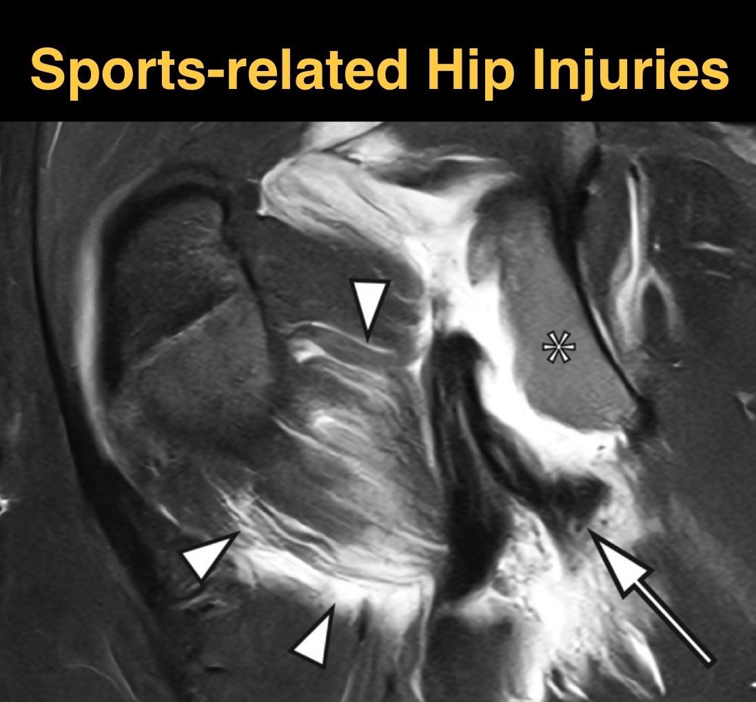
Proximal hamstring injuries. It is common to observe a partial avulsion pattern where the deep tendon fibers detach from the ischial tuberosity while the superficial components, including extensions toward the sacrotuberous ligament, remain continuous. This anatomical

Gonzalo Serrano-Belmar. (@gserranob_msk) 's Twitter Profile Photo

Achilles tendon rupture most commonly occurs in the mid-substance region, approximately 2–6 cm proximal to the calcaneal insertion, where the tendon is relatively hypovascular and often shows underlying tendinosis. On ultrasound, a hypoechoic or anechoic gap may be seen

RadioGraphics (@radiographics) 's Twitter Profile Photo

Let’s learn about radiologic markers, dynamic imaging techniques, & current challenges in establishing diagnostic criteria for hip microinstability. bit.ly/3GC78kp

Let’s learn about radiologic markers, dynamic imaging techniques, &amp; current challenges in establishing diagnostic criteria for hip microinstability. bit.ly/3GC78kp
Reto Sutter (@sutterbalgrist) 's Twitter Profile Photo

#JustPublished: Unicompartmental Knee Arthroplasty (#UKA): What are the Radiographic Predictors for #Conversion to Total Knee Arthroplasty (#TKA)? 👉 Access the full article here (#OpenAccess): doi.org/10.1016/j.ejra… Balgrist University Hospital Balgrist Campus #MSKrad

#JustPublished: Unicompartmental Knee Arthroplasty (#UKA): What are the Radiographic Predictors for #Conversion to Total Knee Arthroplasty (#TKA)?

👉 Access the full article here (#OpenAccess): doi.org/10.1016/j.ejra…

<a href="/derbalgrist/">Balgrist University Hospital</a> <a href="/BalgristCampus/">Balgrist Campus</a> #MSKrad
Sameer Raniga (@samrad77) 's Twitter Profile Photo

Nothing fancy today! Pearl: Elevated pronator quadratus fat plane can be the first clue to a distal radius fracture. Pitfall: Don’t dismiss a “normal” X-ray if the fat plane looks off. Wisdom: Soft tissues often speak before the bone does. —Pearls, pitfalls, and wisdom from

Nothing fancy today! 

Pearl: Elevated pronator quadratus fat plane can be the first clue to a distal radius fracture.

Pitfall: Don’t dismiss a “normal” X-ray if the fat plane looks off.

Wisdom: Soft tissues often speak before the bone does.

—Pearls, pitfalls, and wisdom from
sergio serrano belmar (@sserranobmsk) 's Twitter Profile Photo

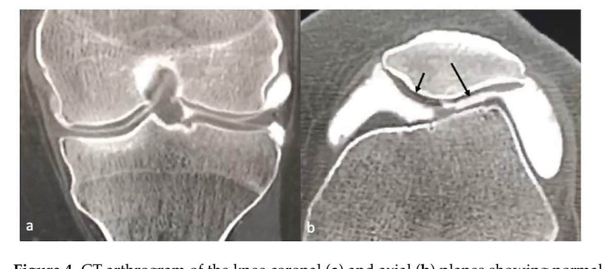
Imaging of Cartilage and Chondral Defects: An Overview doi.org/10.3390/life13… This review article provides an overview of cartilage and chondral defect imaging, emphasizing the role of different modalities in evaluating the health and integrity of articular cartilage. While

Imaging of Cartilage and Chondral Defects: An Overview
doi.org/10.3390/life13…

This review article provides an overview of cartilage and chondral defect imaging, emphasizing the role of different modalities in evaluating the health and integrity of articular cartilage. While
sergio serrano belmar (@sserranobmsk) 's Twitter Profile Photo

Subacute-Chronic Septic Arthritis: Diagnosis by MRI Septic arthritis is a rare complication of knee arthroscopy, with devastating effects if not diagnosed early and treated appropriately. It is clinically diagnosed by progressive, excessive pain for the time following surgery,