Mario Prieto MD PhD (@dr_mprieto) 's Twitter Profile
Mario Prieto MD PhD

@dr_mprieto

Pathologist at Sanchinarro Hospital.
Special interest in #hemepath #breastpath #lymphoma #gynpath #pathology #Molpath

ID: 833770799790247936

calendar_today20-02-2017 20:10:24

6,6K Tweet

3,3K Followers

980 Following

Siba El Hussein, MD (@sibaelhussein) 's Twitter Profile Photo

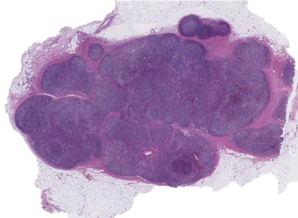
Illustration of the danger of inadvertently sampling germinal centers in limited core-needle biopsies, particularly in the setting of lymphadenopathy with follicular hyperplasia➡️area circled in red with large centroblasts, is an enlarged germinal center (with dark and light

Illustration of the danger of inadvertently sampling germinal centers in limited core-needle biopsies, particularly in the setting of lymphadenopathy with follicular hyperplasia➡️area circled in red with large centroblasts, is an enlarged germinal center (with dark and light
PaulYoungMD (@paulyoungmd1) 's Twitter Profile Photo

Please join our Lymphoma Unknown Conference with Dr Medeiros tomorrow 04/11/25 4pm CST. Link to preview slides: pathpresenter.net/public/present… mdacc.zoom.us/j/89316803503?… Kirill Lyapichev Siba El Hussein, MD SHUYU E MD, PhD Aakash Bhatia, MD Xenia Parisi, MD

Please join our Lymphoma Unknown Conference with Dr Medeiros tomorrow 04/11/25 
4pm CST. 
Link to preview slides: pathpresenter.net/public/present…

mdacc.zoom.us/j/89316803503?…

<a href="/KirillLyapichev/">Kirill Lyapichev</a> 
<a href="/SibaElHussein/">Siba El Hussein, MD</a>
<a href="/EShuyu/">SHUYU E MD, PhD</a>
<a href="/SkybhatiaMD/">Aakash Bhatia, MD</a> 
<a href="/XeniaParisi/">Xenia Parisi, MD</a>
Siba El Hussein, MD (@sibaelhussein) 's Twitter Profile Photo

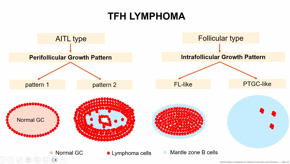
Great cartoon by Dr. Min Shi today depicting the different types of infiltration by TFH lymphoma: 1) Peri-follicular (with patterns 1 and 2) 2) Intra-follicular: a) FL-like, colonizing germinal centers or b) PTGC-like with complete disruption of germinal

Great cartoon by Dr. Min Shi today depicting the different types of infiltration by TFH lymphoma:

1) Peri-follicular (with patterns 1 and 2) 

2) Intra-follicular:

     a) FL-like, colonizing germinal centers 

or 

      b) PTGC-like with complete disruption of germinal
Sanam Loghavi, MD صنم لغوی 🔬🧬 (@sanamloghavi) 's Twitter Profile Photo

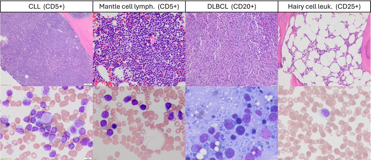
If B cell lymphomas had a calendar today 📆 … 05/05/20 25  😉 a #hemepath #CincoDeMayo #MetGala post 📍CLL (CD5) 📍Mantle cell lymphoma (CD5) 📍DLBCL (CD20) 📍Hairy cell leukemia (CD25) #Nerdy…. I admit 🤓 even for a pathologist! 🙈 #IYKYK

If B cell lymphomas had a calendar today 📆 … 05/05/20 25  😉
a #hemepath #CincoDeMayo #MetGala post
📍CLL (CD5)
📍Mantle cell lymphoma (CD5)
📍DLBCL (CD20)
📍Hairy cell leukemia (CD25)
#Nerdy…. I admit 🤓 even for a pathologist! 🙈
#IYKYK
Siba El Hussein, MD (@sibaelhussein) 's Twitter Profile Photo

Micronodular T-cell/histiocyte-rich large B-cell lymphoma (MTLBL) is unique variant of T-cell/histiocyte-rich large B-cell lymphoma, characterized by primary splenic presentation #Hemepath #PathX #PathTwitter #Lymsm #Surgpath #MedEd #GIpath #SpleenPath

Micronodular T-cell/histiocyte-rich large B-cell lymphoma (MTLBL) is unique variant of T-cell/histiocyte-rich large B-cell lymphoma, characterized by primary splenic presentation 

#Hemepath #PathX #PathTwitter #Lymsm #Surgpath #MedEd #GIpath #SpleenPath
Cristina D. del Arco (@crisdelarco) 's Twitter Profile Photo

Maravillosa la charla sobre HER2 en mama de Laura Comerma en el congreso #SEAP2025SS. Ha incluido en su presentación una carta/crítica que merece la pena leer (DOI: 10.1056/NEJMc2416654) 📚

Maravillosa la charla sobre HER2 en mama de Laura Comerma en el congreso #SEAP2025SS. Ha incluido en su presentación una carta/crítica que merece la pena leer (DOI: 10.1056/NEJMc2416654) 📚
Rumeal D. Whaley, MD (@rdwhaleymd) 's Twitter Profile Photo

Read our latest #research in Human Pathology Evaluation of 3,606 renal cell tumors for TFE3 rearrangements and TFEB alterations via FISH, NGS, and GPNMB IHC Free access until July 12, 2025. kwnsfk27.r.eu-west-1.awstrack.me/L0/https:%2F%2… #GUPath
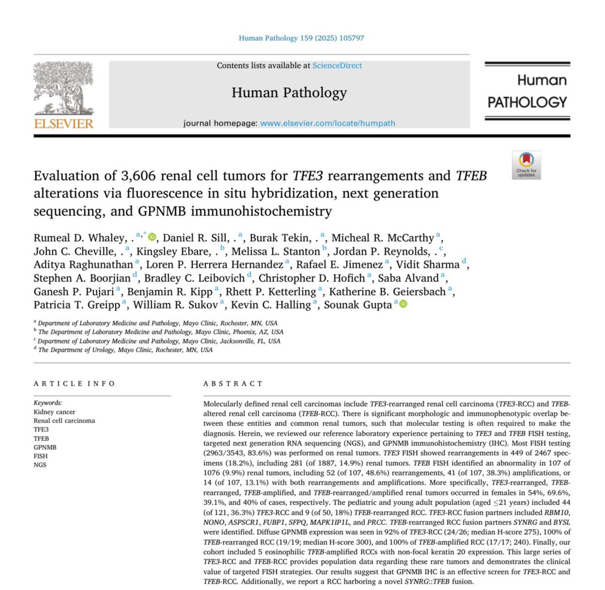
Read our latest #research in <a href="/Human_Pathology/">Human Pathology</a> 

Evaluation of 3,606 renal cell tumors for TFE3 rearrangements and TFEB alterations via FISH, NGS, and GPNMB IHC

Free access until July 12, 2025. 

kwnsfk27.r.eu-west-1.awstrack.me/L0/https:%2F%2…

#GUPath
Eduardo Alcaraz, MD PhD (@edusqo) 's Twitter Profile Photo

Enhorabuena a l@s premiados en #SEAP2025SS SEAP-IAP y sobre todo a los Drs. Matias-Guiu Xavier Matias-Guiu G, Marcial García Rojo Patologia Digital, Ignacio Ruz-Caracuel Ignacio Ruz-Caracuel y Sara Quiñones Sara Quiñones. Muy merecido 👏🏾 🙌🏾 ♥️

Enhorabuena a l@s premiados en #SEAP2025SS <a href="/SEAP_IAP/">SEAP-IAP</a> y sobre todo a los Drs. Matias-Guiu <a href="/xmatiasguiu/">Xavier Matias-Guiu G</a>, Marcial García Rojo <a href="/PatologoDigital/">Patologia Digital</a>, Ignacio Ruz-Caracuel <a href="/PathRuzCaracuel/">Ignacio Ruz-Caracuel</a> y Sara Quiñones <a href="/draquinnones/">Sara Quiñones</a>.
Muy merecido 👏🏾 🙌🏾 ♥️
Fatima Shamsuddin (@fatimashamspath) 's Twitter Profile Photo

Grateful to the Almighty, my mentors, colleagues, and family. Article titled — “FH Deficient Uterine Leiomyomas: Pathological Insights into a Rare Tumor” — has been accepted for publication in the Gulf Journal of Oncology, Issue 48, May 2025. Article link:

Grateful to the Almighty, my mentors, colleagues, and family. Article titled — “FH Deficient Uterine Leiomyomas: Pathological Insights into a Rare Tumor” — has been accepted for publication in the Gulf Journal of Oncology, Issue 48, May 2025. 
Article link:
NotasDePatologia (@notas_patologia) 's Twitter Profile Photo

🧬 Castleman Disease – Hyaline Vascular Variant (HVCD): a vascular spectacle in lymph nodes! Castleman disease is a benign lymphoproliferative disorder that can present in a unicentric or multicentric form. Histologically, it is classified into two main types: the plasma cell

🧬 Castleman Disease – Hyaline Vascular Variant (HVCD): a vascular spectacle in lymph nodes!

Castleman disease is a benign lymphoproliferative disorder that can present in a unicentric or multicentric form. Histologically, it is classified into two main types: the plasma cell
SEAP-IAP (@seap_iap) 's Twitter Profile Photo

¡Mañana es el día! 🎉 A las 14:00h (GMT+1) conectamos con el webinar #HER2low 🧬 La Dra. Pérez Mies Belen Perez Mies compartirá resultados y casos clave de las rondas de calidad SEAP 🧠📝 🆓 Inscripción gratuita para socios en 👉 seap.es ¡No te lo pierdas!

¡Mañana es el día! 🎉
A las 14:00h (GMT+1) conectamos con el webinar #HER2low 🧬
La Dra. Pérez Mies <a href="/Bepemie/">Belen Perez Mies</a> compartirá resultados y casos clave de las rondas de calidad SEAP 🧠📝
🆓 Inscripción gratuita para socios en 👉 seap.es
¡No te lo pierdas!

Janira Navarro (@janiranavarro) 's Twitter Profile Photo

HER2 IHC endometrium reporting template Endometrium (USC): Use both endometrium HER2 scoring (Dr. Buza/Fader scoring) and gastric HER2 scoring Dr. Troxell (information courtesy Dr. McHenry/Stanford GYN path) #USCPAP25 #pathology #PathX

HER2 IHC endometrium reporting template
Endometrium (USC): Use both endometrium HER2 scoring (Dr. Buza/Fader scoring) and gastric HER2 scoring

Dr. Troxell (information courtesy Dr. McHenry/Stanford GYN path) #USCPAP25 #pathology #PathX
Human Pathology (@human_pathology) 's Twitter Profile Photo

New in #HumPathol: Current understanding of phyllodes tumors of the breast: Tumor classification, molecular landscape, and best pathology practice. sciencedirect.com/science/articl… #pathology #PathTwitter #PathX #breastpath

New in #HumPathol: Current understanding of phyllodes tumors of the breast: Tumor classification, molecular landscape, and best pathology practice. sciencedirect.com/science/articl… #pathology #PathTwitter #PathX #breastpath
Curtis Lachowiez (@clachowiez) 's Twitter Profile Photo

Happy to share our newest work out today in BCD evaluating disease classification and prognosis in the “world of myeloid malignancies”! Wonderful collaboration with Sanam Loghavi, MD صنم لغوی 🔬🧬 Elli Papaemmanuil, PhD Elsa Bernard and colleagues.

Muhammad Hussain, MD (@whosainastro) 's Twitter Profile Photo

2/ Ki-67 was originally identified as an antigen recognized by a monoclonal antibody, created by immunizing mice with nuclei isolated from the Hodgkin lymphoma cell line L428.

Muhammad Hussain, MD (@whosainastro) 's Twitter Profile Photo

4/ Ki-67 was first described in the early 1980s by Dr. Johannes Gerdes and colleagues at the University of Kiel. They were searching for proteins that are expressed only in actively dividing cells, a potential marker of cell proliferation.