Jay R. Seltzer (@jrseltzer) 's Twitter Profile
Jay R. Seltzer

@jrseltzer

Medical Director - Urine Microscopy Laboratory Missouri Baptist Medical Center - St. Louis MO

ID: 238010563

linkhttp://stlkidney.com calendar_today14-01-2011 04:28:52

5,5K Tweet

5,5K Followers

566 Following

Jay R. Seltzer (@jrseltzer) 's Twitter Profile Photo

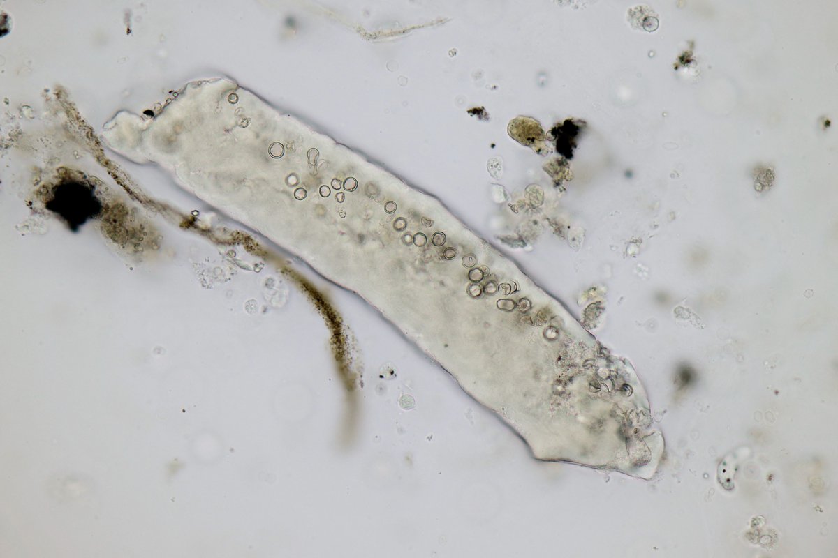
Calcium phosphate crystals - brightfield and darkfield illumination, unstained - in the urine of a patient with MPA - #UrineMicroscopy - Could be the new X logo! Elon Musk

Calcium phosphate crystals - brightfield and darkfield illumination, unstained -  in the urine of a patient with MPA - #UrineMicroscopy -  Could be the new X logo!
<a href="/elonmusk/">Elon Musk</a>
Jay R. Seltzer (@jrseltzer) 's Twitter Profile Photo

WBC cast - brightfield with SM stain - from patient with recurrent UTI's s/p abx rx over 3 weeks - #UrineMicroscopy #UrinarySediment

WBC cast - brightfield with SM stain - from patient with recurrent UTI's s/p abx rx over 3 weeks - #UrineMicroscopy #UrinarySediment
Jay R. Seltzer (@jrseltzer) 's Twitter Profile Photo

Vacation's over and I'm back in the lab.... Young male presented with DVT and PE. Found to have UPC 8 gm/gm and serum albumin 2.2 and creatinine 2.3 - #UrineMicroscopy showed RBC casts with fibrinous matrix as well as a few lipid casts - brightfield with SM stain -

Vacation's over and I'm back in the lab....
Young male  presented with DVT and PE. Found to have UPC 8 gm/gm and serum albumin 2.2 and creatinine 2.3 - #UrineMicroscopy showed RBC casts with fibrinous matrix as well as a few lipid casts - brightfield with SM stain -
Jay R. Seltzer (@jrseltzer) 's Twitter Profile Photo

Young male presented with DVT and PE. Found to have UPC 8 gm/gm, serum albumin 2.2, and creatinine 2.3 - #UrineMicroscopy showed this mixed cellular cast containing RBC's, tubular epithelial cells, and WBC's - brightfield with SM stain - #UrinarySediment

Young male  presented with DVT and PE. Found to have UPC 8 gm/gm, serum albumin 2.2, and creatinine 2.3 - #UrineMicroscopy showed this mixed cellular cast containing RBC's, tubular epithelial cells, and WBC's - brightfield with SM stain - #UrinarySediment
Jay R. Seltzer (@jrseltzer) 's Twitter Profile Photo

New #UrinarySediment and #UrineMicroscopy case studies in AKDH Use the link below but be sure to view as a pdf - the online version is not formatted correctly kwnsfk27.r.eu-west-1.awstrack.me/L0/https:%2F%2…

New #UrinarySediment  and #UrineMicroscopy case studies in <a href="/akdhjournal/">AKDH</a> 

Use the link below but be sure to view as a pdf - the online version is not formatted correctly

kwnsfk27.r.eu-west-1.awstrack.me/L0/https:%2F%2…
Swetha Kanduri (@swethakandurimd) 's Twitter Profile Photo

Great opportunities to deepen the knowledge on the art of urine microscopy, it’s techniques and clinical utility 🌟Come join us for a hands on work shop with experts in the field at KIDNEYcon May 1-3 2025 Juan Carlos Q Velez Jay R. Seltzer kidneycon.org/registration

Jay R. Seltzer (@jrseltzer) 's Twitter Profile Photo

Good example of the different appearances of RBC's, WBC's and tubular epithelial cells under phase contrast #UrineMicroscopy

Jay R. Seltzer (@jrseltzer) 's Twitter Profile Photo

Vacuolar cast (which probably also contains some RBC's) - brightfield with SM stain - from patient with AKI and suspected hydralazine-induced vasculitis #UrineMicroscopy #UrinarySediment

Vacuolar cast (which probably also contains some RBC's) - brightfield with SM stain - from patient with AKI and suspected hydralazine-induced vasculitis #UrineMicroscopy #UrinarySediment
Jay R. Seltzer (@jrseltzer) 's Twitter Profile Photo

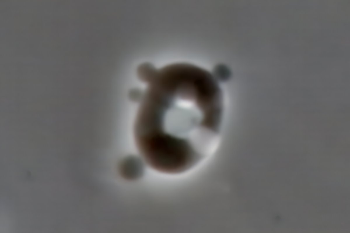
Acanthocyte - darkfield, unstained - cropped from original x1000 - from case of suspected hydralazine-induced vasculitis - #UrineMicroscopy #UrinarySediment

Acanthocyte - darkfield, unstained - cropped from original x1000 - from case of suspected hydralazine-induced vasculitis - #UrineMicroscopy #UrinarySediment
Jay R. Seltzer (@jrseltzer) 's Twitter Profile Photo

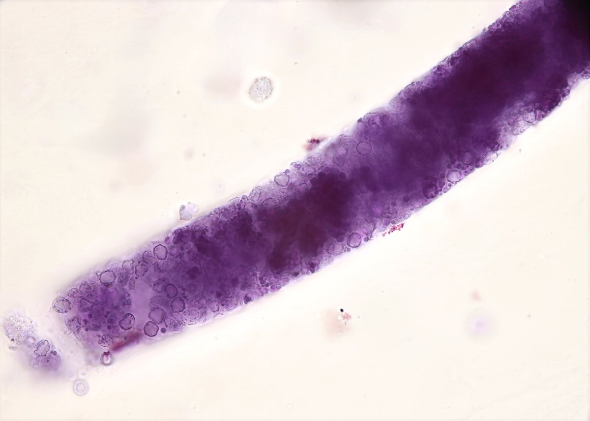
RBC casts - brightfield with SM stain - from case of suspected hydralazine-induced vasculitis - #UrineMicroscopy #UrinarySediment

RBC casts - brightfield with SM stain - from case of suspected hydralazine-induced vasculitis - #UrineMicroscopy #UrinarySediment