Ulf Ahlgren´s Research Group (Umeå University)
@lighttodiabetes
By bringing light to diabetes by optical (mesoscopic) imaging, our research aims at increasing our understanding of the pathophysiology of diabetes.
ID: 1186949199138607105
https://www.umu.se/en/research/groups/ulf-ahlgren2/?languageId=1 23-10-2019 10:16:13
22 Tweet
49 Followers
68 Following
 's Twitter Profile Photo")
 on Twitter photo 🔥The 9th Round of Easy Loan, Earn $40 Reward is in progress❗️
⏰ Promotion Period: January 15th - Feburary 15th, 2025
👉 Register now and check more details at gate.io/campaigns/358")
 (@lighttodiabetes) on Twitter photo On world diabetes day we would like to raise awareness of the need for diabetes research and thank our funders for their support.")
 (@lighttodiabetes) on Twitter photo Bringing light to diabetes on world diabetes day: 6 Lasers are used to analyse a pancreatic specimen in a Lavision Biotec 2nd generation Ultramicroscope.")
 (@lighttodiabetes) on Twitter photo 🔬Check out this stunning fluorescent slab of human pancreas in our 3D Microscope (Optical Projection Tomography). We make the sample transparent by adapting the refractive index, which allows to shine through the sample.")
Now published! A new study in Nature's Communications Biology on the Streptozotocin diabetes mouse model, the most used diabetes model in rodents. Using advanced imaging, we show that the majority of islets remain, but surviving islets of Langerhans are dysfunctional. nature.com/articles/s4200…
 (@umeafor) 's Twitter Profile Photo")
 (@umeafor) on Twitter photo Breaking news! UCMM would like to congratulate Prof. Ulf Ahlgren’s group on their excellent new paper published yesterday in Communications Biology (nature.com/articles/s4200…). The image depicts a mouse pancreas imaged by Optical Projection Tomography (OPT).")
 's Twitter Profile Photo")
 's Twitter Profile Photo")
 on Twitter photo 🔎 Researchers from @UmeaUniversity show that hyperglycemic #Streptozotocin-treated mice still harbor a large pool of remaining β-cells but display pancreas-wide downregulation of glucose transporter type 2.
🔓 Open access go.nature.com/34zGkKT")
 's Twitter Profile Photo")
 on Twitter photo Congratulations Professor Emmanuelle Charpentier for the award of the 2020 Nobel Prize in Chemistry for the discovery of the gene-editing tool CRISPR-Cas9.
#EmmanuelleCharpentier #NobelPrize
umu.se/en/news/discov…")
 's Twitter Profile Photo")
 's Twitter Profile Photo")
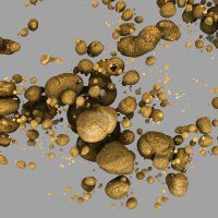
Reconstruction of the 3 dimensional anatomy of the human endocrine pancreas #islets Communications Biology nature.com/articles/s4200…
 on Twitter photo Reconstruction of the 3 dimensional anatomy of the human endocrine pancreas #islets <a href=\"/CommsBio/\">Communications Biology</a> nature.com/articles/s4200…")
Our article is out Communications Biology! By the use of a 3D printed matrix we show how human organs (in our case the pancreas) can be specifically labelled, imaged by optical mesoscopic imaging techniques & reconstructed back together in 3D. nature.com/articles/s4200… #LSFM #imaging Umea Centre for Molecular Medicine (UCMM)
 's Twitter Profile Photo")
 on Twitter photo Ny metod möjliggör detaljstudier av mänskliga organ i 3D mynewsdesk.com/se/diabeteswel…")
In this study, Hahn et al. present a method for visualizing the endocrine human pancreas in 3D and apply it to individuals with type 2 diabetes to reveal morphological differences in the endocrine pancreas. Ulf Ahlgren´s Research Group (Umeå University) Umea Centre for Molecular Medicine (UCMM) nature.com/articles/s4200…
 on Twitter photo In this study, Hahn et al. present a method for visualizing the endocrine human pancreas in 3D and apply it to individuals with type 2 diabetes to reveal morphological differences in the endocrine pancreas. <a href=\"/lighttodiabetes/\">Ulf Ahlgren´s Research Group (Umeå University)</a> <a href=\"/UmeaFor/\">Umea Centre for Molecular Medicine (UCMM)</a> nature.com/articles/s4200…")
 's Twitter Profile Photo")
 's Twitter Profile Photo")
After a difficult time, we’re back on track and our 3rd manuscript of the year has been accepted for publication! 😀 A great collaboration with Ulf Ahlgren´s Research Group (Umeå University) & Peter Thorn “Insulin-binding peptide probes provide a novel strategy for pancreatic β-cell imaging” ACS Publications Bio & Med Chem Content
 's Twitter Profile Photo")
Individual islets reconstructed into the 3D #pancreas using a novel imaging approach. #WorldDiabetesDay2021 #WorldDiabetesDay Video and research by Max Hahn et al Ulf Ahlgren´s Research Group (Umeå University) @UmeaUniversity in Communications Biology. Read more on bpod.mrc.ac.uk/archive/2021/1… with @MRV_LMS and John Ankers
We made the cover! Insulin-Binding Peptide Probes Provide a Novel Strategy for Pancreatic β-Cell Imaging. ULB ComRecherche NCP F.R.S.-FNRS Diabetes Australia JDRF Research #EFSD. Great collaboration with Ulf Ahlgren´s Research Group (Umeå University) and Peter Thorn pubs.acs.org/doi/10.1021/ac…
 on Twitter photo We made the cover! Insulin-Binding Peptide Probes Provide a Novel Strategy for Pancreatic β-Cell Imaging. <a href=\"/ULBRecherche/\">ULB ComRecherche</a> <a href=\"/NCP_frsFNRS/\">NCP F.R.S.-FNRS</a> <a href=\"/DiabetesAus/\">Diabetes Australia</a> <a href=\"/JDRFResearch/\">JDRF Research</a> #EFSD. Great collaboration with <a href=\"/lighttodiabetes/\">Ulf Ahlgren´s Research Group (Umeå University)</a> and <a href=\"/pthorn14/\">Peter Thorn</a>
pubs.acs.org/doi/10.1021/ac…")