Maria Martinez-Lage, MD (she/her/ella) 🏳️‍🌈 (@mlage) 's Twitter Profile
Maria Martinez-Lage, MD (she/her/ella) 🏳️‍🌈

@mlage

Neuropathologist, Assistant Prof, Program Director @mgbpathology Trained @PennPathLabMed. From Pamplona, Spain #Neuropath #Pathology #antiRacism

ID: 12382182

calendar_today18-01-2008 01:41:35

4,4K Tweet

4,4K Followers

2,2K Following
Mass General Brigham Pathology (@mgbpathology) 's Twitter Profile Photo

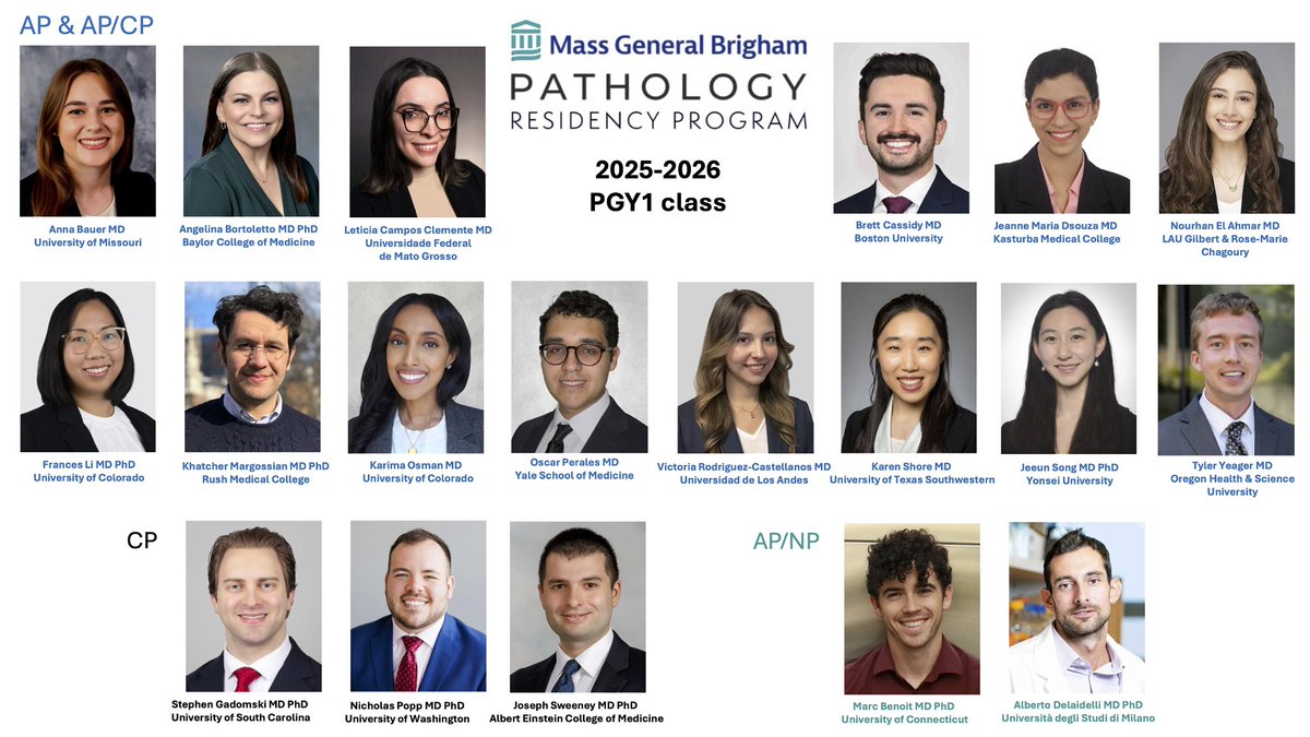
We couldn’t be more thrilled to introduce this stellar new class of 19 (yes, 19!) PGY1s joining the Mass General Brigham pathology family! 🥳 Welcome to the team — we can’t wait to have you on board and see you thrive! 🧬🔬💫

We couldn’t be more thrilled to introduce this stellar new class of 19 (yes, 19!) PGY1s joining the Mass General Brigham pathology family! 🥳
Welcome to the team — we can’t wait to have you on board and see you thrive! 🧬🔬💫
Maria Martinez-Lage, MD (she/her/ella) 🏳️‍🌈 (@mlage) 's Twitter Profile Photo

Welcome to this fantastic class and congratulations!! Enjoy some well deserved rest now before the fun begins in the summer. 🤓🎉 #path2path #pathmatch2025 #pathmatch

Mass General Brigham Pathology (@mgbpathology) 's Twitter Profile Photo

Some of our amazing team representing at #USCAP2025! Proud to see their hard work and brilliant science on display. Doing what we do best: science and style. 😉

Some of our amazing team representing at #USCAP2025! Proud to see their hard work and brilliant science on display. Doing what we do best: science and style. 😉
Mireille Bitar, M.D. (@bitar_mireille) 's Twitter Profile Photo

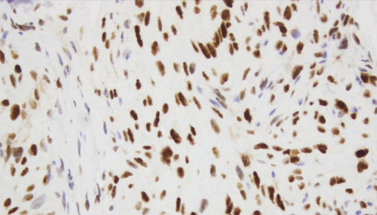
History of spine radiation as a child for a CNS neoplasm, few years later with a spinal bone lesion (H&E and p53 IHC).. no osteoid formation seen in the tumor, likely diagnosis? #neuropath #pedipath #molecpath #bonepath

History of spine radiation as a child for a CNS neoplasm, few years later with a spinal bone lesion (H&E and p53 IHC).. no osteoid formation seen in the tumor,  likely diagnosis? #neuropath #pedipath #molecpath #bonepath
AANP (@neuropathology) 's Twitter Profile Photo

Don’t forget! AANP Teaching Rounds on Monday, March 31 from 11am-12pm EST: Anti-Amyloid Beta Immunotherapy-Related Vasculitis by Dr. Rudy Castellani. More information here: neuropath.org/teaching-rounds #neuropath #neuropathology #pathology

Laura Molina Porcel 🧠 🩺🔬⛰️ (@lauramolinapor1) 's Twitter Profile Photo

What a morning! Presenting the results of the study on the correlation between genetic risk variants for AD and PD and neuropathological findings of 831 patients from the Neurological Tissue Bank of Hospital Clínic IDIBAPS in collaboration with University of Miami, at the #ADPDMeeting.

What a morning! Presenting the results of the study on the correlation between genetic risk variants for AD and PD and neuropathological findings of 831 patients from the Neurological Tissue Bank of <a href="/hospitalclinic/">Hospital Clínic</a> <a href="/idibaps/">IDIBAPS</a> in collaboration with <a href="/univmiami/">University of Miami</a>, at the #ADPDMeeting.
Jared T. Ahrendsen MD, PhD (AP/NP/FP) (@jaredahrendsen) 's Twitter Profile Photo

Cerebellar #brain mass from a child. Can you render a #diagnosis, including most likely molecular classification, with only these lower power H&E and reticulin images? #neuropath 🧠🔬💪

Cerebellar #brain mass from a child. Can you render a #diagnosis, including most likely molecular classification, with only these lower power H&amp;E and reticulin images? 
#neuropath 🧠🔬💪
Craig Horbinski (@craighorbinski) 's Twitter Profile Photo

24 year-old woman with a posterior fossa mass. NGS picked up an FGFR1 mutation. Diagnosis? (As #tolkien might have said, the Rays of Fëanor Gleam 'Neath Telperion while you contemplate this one.) #pathology #neuropath #pathtwitter

24 year-old woman with a posterior fossa mass. NGS picked up an FGFR1 mutation. Diagnosis? (As #tolkien might have said, the Rays of Fëanor Gleam 'Neath Telperion while you contemplate this one.) #pathology #neuropath #pathtwitter
AANP (@neuropathology) 's Twitter Profile Photo

Diffuse astrocytoma, MYB or MYBL1-altered. An infiltrating, but grade 1 glioma! (Note the trapped neurons—evidence of infiltration) Tumor cells rel. monomorphic, round to ovoid or spindly. It is strongly associated with epilepsy. #neuropath #neuropathology #pathx #PathTwitter

Diffuse astrocytoma, MYB or MYBL1-altered. An infiltrating, but grade 1 glioma! (Note the trapped neurons—evidence of infiltration) Tumor cells rel. monomorphic, round to ovoid or spindly.  It is strongly associated with epilepsy.  #neuropath #neuropathology #pathx #PathTwitter
AANP (@neuropathology) 's Twitter Profile Photo

A similar tumor, with molecular overlap and also grade 1 is angiocentric glioma. This tumor can have MYB-Astro like areas; much more characteristic is bland spindle cells arranged angiocentrically. #neuropath #neuropathology #cnspathology #pathx #pathtwitter

A similar tumor, with molecular overlap and also grade 1 is angiocentric glioma.  This tumor can have MYB-Astro like areas; much more characteristic is bland spindle cells arranged angiocentrically. #neuropath #neuropathology #cnspathology #pathx #pathtwitter
Jay Hwang (@path4people) 's Twitter Profile Photo

Cerebellar Tumor in a 10 year old 🧒 👧 🧬 Molecular shows KIAA1549-BRAF fusion Cyto ⬇️ Dx? 🤔 #neurosurgery #pathx #pathology #neuropath #radiology

Cerebellar Tumor in a 10 year old 🧒 👧 

🧬 Molecular shows KIAA1549-BRAF fusion

Cyto ⬇️  Dx?  🤔 

#neurosurgery #pathx #pathology #neuropath #radiology
Kenneth Aldape, MD (@kenneth_aldape) 's Twitter Profile Photo

Methylation Case of the Week: 3 young-middle aged adults with lesions in the cerebral hemisphere of the same tumor type. Chromosome 13 loss in all 3 was found. Any thoughts on what this might be! just think How Proud And Privileged you will feel to make the diagnosis!#pathology

Methylation Case of the Week:

3 young-middle aged adults with lesions in the cerebral hemisphere of the same tumor type. Chromosome 13 loss in all 3 was found. Any thoughts on what this might be! just think How Proud And Privileged you will feel to make the diagnosis!#pathology
Brain Pathology (@brainpathol) 's Twitter Profile Photo

cIMPACT-NOW update 10: Recommendations for defining new types for central nervous system tumor classification #neuropath Kenneth Aldape, MD Sahm Lab Hawkins Lab onlinelibrary.wiley.com/doi/10.1111/bp…

Esther Youd (@estheryoud) 's Twitter Profile Photo

Next RCPath autopsy webinar: Neuropathology for the non-specialist Weds 11th June 1-2pm BST Dr Robin Highley #autopsy #neuropath #forensicpath Register below: rcpath.org/event/autopsy-…

Karleen Meiklejohn, MD 💀💉🔬 (@drkmeiklejohn) 's Twitter Profile Photo

Never a bad time to consider these things, plus there's some good job market discussion! link.springer.com/article/10.100… #PathTwitter #pathology

Never a bad time to consider these things, plus there's some good job market discussion!
link.springer.com/article/10.100… #PathTwitter #pathology
Kenneth Aldape, MD (@kenneth_aldape) 's Twitter Profile Photo

Methylation Case of the Week: 3 year old female patient with a intramedullary (C7 to T8) lesion. The diagnostic fusion was found. You won't need to believe in ASTROlogy to have a BLAST trying to diagnose this case. #pathology #neuropath #Pathtwitter

Methylation Case of the Week:

3 year old female patient with a intramedullary (C7 to T8) lesion. The diagnostic fusion was found. You won't need to believe in ASTROlogy to have a BLAST trying to diagnose this case. #pathology #neuropath #Pathtwitter
Mireille Bitar, M.D. (@bitar_mireille) 's Twitter Profile Photo

Brain mass in a young patient suspected to be a vascular malformation based on imaging..what immunostains to get? #pedipath #neuropath

Brain mass in a young patient suspected to be a vascular malformation based on imaging..what immunostains to get?
#pedipath #neuropath
Silvia Judith Hernández (@silvia_hdzmd) 's Twitter Profile Photo

4 Cytologic Key Features in Embryonal tumors during Intra-op consult: 1. Cellular Arrangement: Dyscohesive 2. Background: Scarce/Necrosis 3. Cytoplasm: Scarce 4. Nucleus: Round/Oval, hyperchromatic, pleomorphic, mitosis, indented borders #Neuropath

4 Cytologic Key Features in Embryonal tumors during Intra-op consult:
1. Cellular Arrangement: Dyscohesive
2. Background: Scarce/Necrosis 
3. Cytoplasm: Scarce
4. Nucleus: Round/Oval, hyperchromatic, pleomorphic, mitosis, indented borders #Neuropath