Neurosurgical Atlas (@neurosurgatlas) 's Twitter Profile
Neurosurgical Atlas

@neurosurgatlas

Dr. Aaron Cohen-Gadol’s most respected & popular collection of advanced microneurosurgical techniques in the world. linktr.ee/NeurosurgicalA…

ID: 787020506

linkhttps://www.neurosurgicalatlas.com/ calendar_today28-08-2012 13:32:24

6,6K Tweet

26,26K Followers

825 Following

Neurosurgical Atlas (@neurosurgatlas) 's Twitter Profile Photo

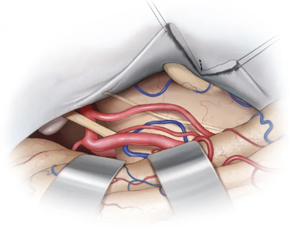
Trochlear nerve should be dissected from its dural canal up to its entrance in the cavernous sinus. The trochlear nerve is free of the tentorial flap, creating a supra- and infratrochlear working window. Learn more here | zurl.co/pUmrz #Neurosurgery

Trochlear nerve should be dissected from its dural canal up to its entrance in the cavernous sinus. The trochlear nerve is free of the tentorial flap, creating a supra- and infratrochlear working window. 

Learn more here | zurl.co/pUmrz 

#Neurosurgery
Neurosurgical Atlas (@neurosurgatlas) 's Twitter Profile Photo

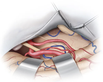
Extension of the tentorial incision up to the Meckel’s cave allow anterolateral reflection of the medial tentorial flap. If the incision extends to the most lateral aspect of the Meckel’s cave, which structures can be visualized? #Neurosurgery #CranialBaseSurgery

Extension of the tentorial incision up to the Meckel’s cave allow anterolateral reflection of the medial tentorial flap. If the incision extends to the most lateral aspect of the Meckel’s cave, which structures can be visualized?  

#Neurosurgery #CranialBaseSurgery
Neurosurgical Atlas (@neurosurgatlas) 's Twitter Profile Photo

If the tentorial incision is extended to the most lateral aspect of the Meckel’s cave, the trigeminal nerve and the origin of the anterior inferior cerebellar artery (AICA) can be visualized. Learn more here: zurl.co/YQyAG #Neurosurgery #CranialBaseSurgery

If the tentorial incision is extended to the most lateral aspect of the  Meckel’s cave, the trigeminal nerve and the origin of the anterior  inferior cerebellar artery (AICA) can be visualized. 

Learn more here: zurl.co/YQyAG 

#Neurosurgery #CranialBaseSurgery
Neurosurgical Atlas (@neurosurgatlas) 's Twitter Profile Photo

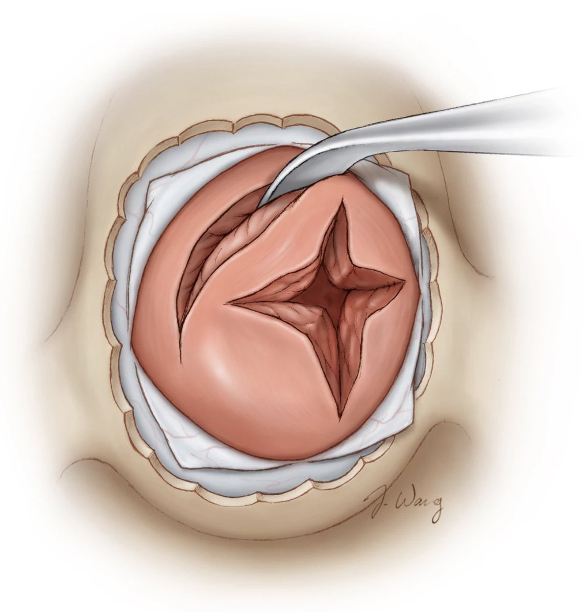
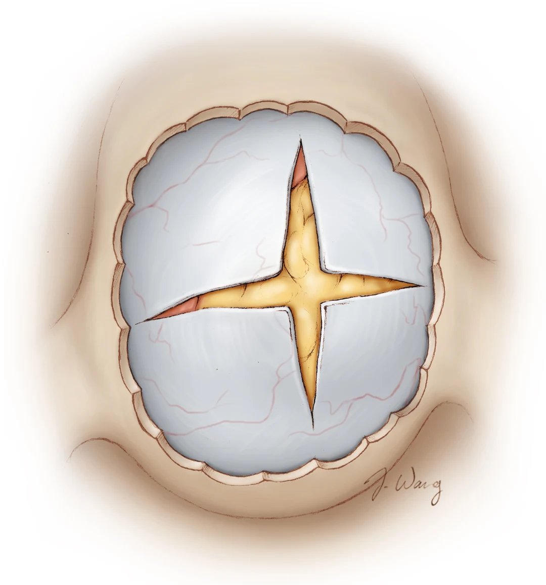
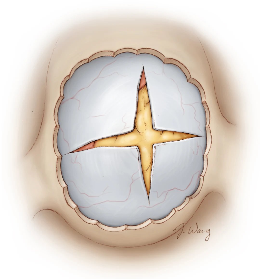
After making a cruciate incision like the one shown here to expose the pituitary gland, what are the key intraoperative features that help distinguish a microadenoma from normal gland tissue? #Neurosurgery #EndoscopicSurgery #PituitaryTumor #Neuroanatomy

After making a cruciate incision like the one shown here to expose the pituitary gland, what are the key intraoperative features that help distinguish a microadenoma from normal gland tissue? 

#Neurosurgery #EndoscopicSurgery #PituitaryTumor #Neuroanatomy
Neurosurgical Atlas (@neurosurgatlas) 's Twitter Profile Photo

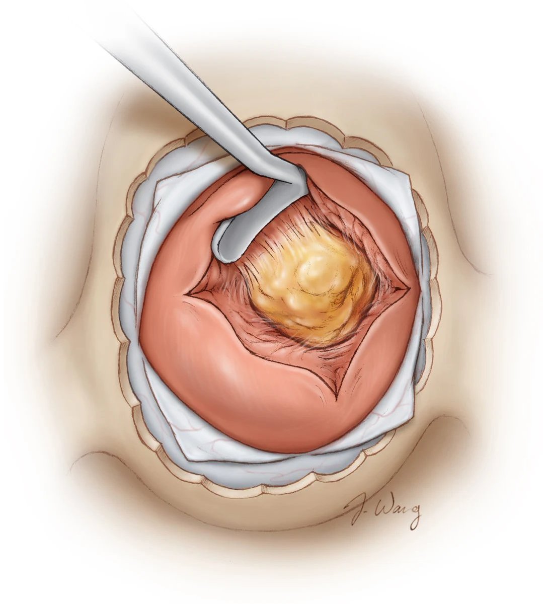
During surgery, a pituitary microadenoma can be distinguished from normal gland tissue by its lighter yellow color, softer texture, and reduced vascularity. It often has a subtle dissection plane separating it from the normal gland. In Cushing’s disease, it’s typically centrally

During surgery, a pituitary microadenoma can be distinguished from normal gland tissue by its lighter yellow color, softer texture, and reduced vascularity. It often has a subtle dissection plane separating it from the normal gland. In Cushing’s disease, it’s typically centrally
Dr. Z Neurosurgery (@drzneurosurgery) 's Twitter Profile Photo

The 3 planes of a brain MRI. To best localize a lesion, a neurosurgeon will look at these three views to triangulate an approach. #Radiology #MRI #Radiologist #Neurosurgeon #Brain #BrainScan #Anatomy #AnatomyAndPhysiology #NeuroAnatomy Neurosurgical Atlas
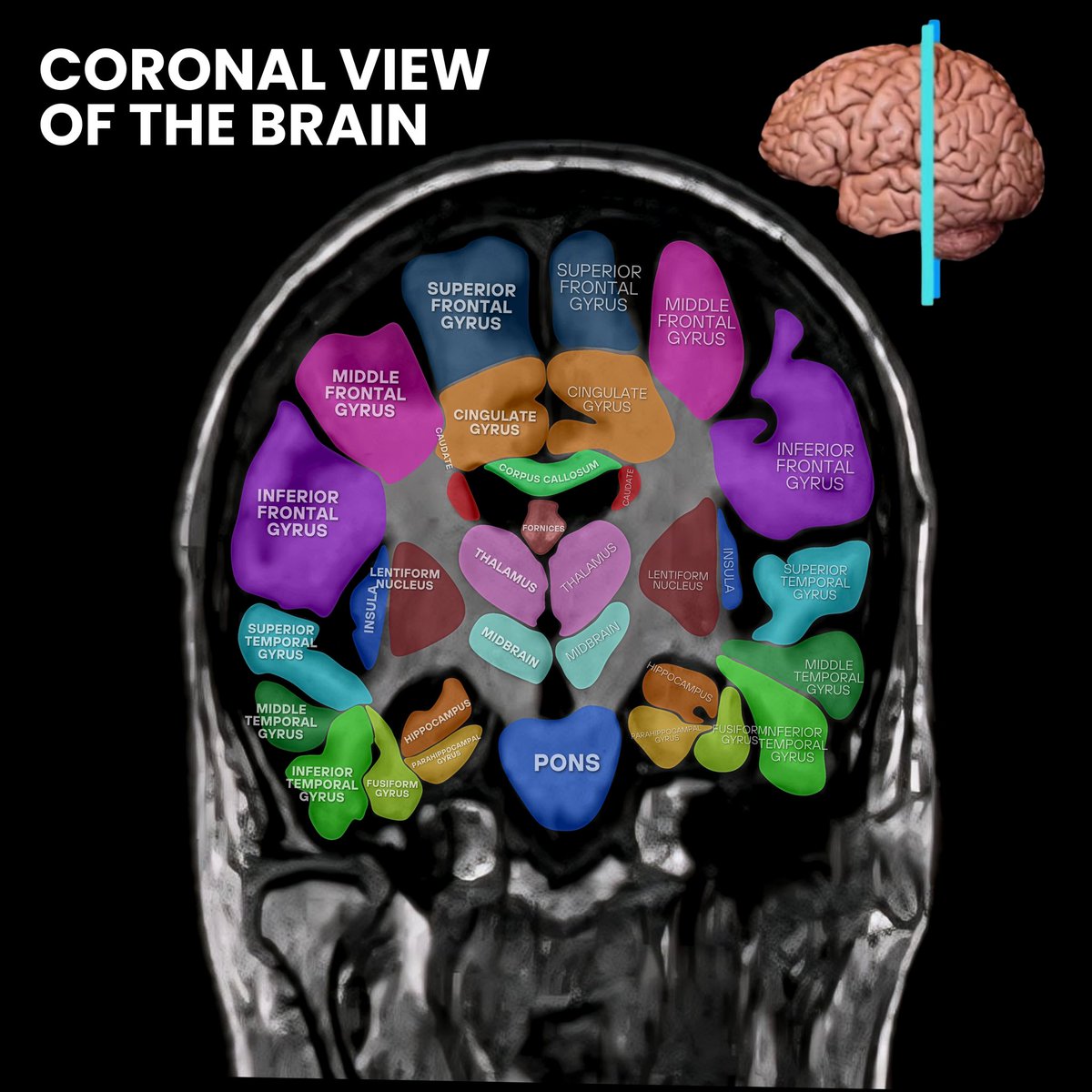
The 3 planes of a brain MRI. To best localize a lesion, a neurosurgeon will look at these three views to triangulate an approach. #Radiology #MRI #Radiologist #Neurosurgeon #Brain #BrainScan #Anatomy #AnatomyAndPhysiology #NeuroAnatomy <a href="/neurosurgatlas/">Neurosurgical Atlas</a>
Neurosurgical Atlas (@neurosurgatlas) 's Twitter Profile Photo

Following tumor removal of a microadenoma for Cushing’s disease, what next step should the surgeon take to improve outcomes? #Neurosurgery #EndoscopicSurgery #PituitaryTumor #Neuroanatomy

Following tumor removal of a microadenoma for Cushing’s disease, what next step should the surgeon take to improve outcomes? 

#Neurosurgery #EndoscopicSurgery #PituitaryTumor #Neuroanatomy
Neurosurgical Atlas (@neurosurgatlas) 's Twitter Profile Photo

In the case of Cushing’s disease and in select cases, the wall of the resection cavity may be removed and pathologically examined. This maneuver may improve the odds of complete endocrine remission. Learn more here: zurl.co/d0m11 #Neurosurgery #EndoscopicSurgery

In the case of Cushing’s disease and in select cases, the wall of the resection cavity may be removed and pathologically examined. This maneuver may improve the odds of complete endocrine remission.

Learn more here: zurl.co/d0m11 

 #Neurosurgery #EndoscopicSurgery
Neurosurgical Atlas (@neurosurgatlas) 's Twitter Profile Photo

In this operative video, Dr. Cohen demonstrates the steps of a temporal craniotomy for resection of a temporal lobe glioblastoma. After scalp incision with Raney clip application, the temporalis muscle was mobilized inferiorly using cautery. Two burr holes were placed, one

Neurosurgical Atlas (@neurosurgatlas) 's Twitter Profile Photo

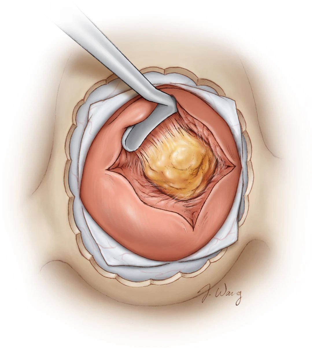
Due to its small size and central location, pituitary microadenomas may be difficult to see. In the case that the tumor is not originally visible, what tool should be used (and in what direction) to get the tumor within the surgical field? #Neurosurgery #EndoscopicSurgery

Due to its small size and central location, pituitary microadenomas may be difficult to see. In the case that the tumor is not originally visible, what tool should be used (and in what direction) to get the tumor within the surgical field? 

#Neurosurgery #EndoscopicSurgery
Neurosurgical Atlas (@neurosurgatlas) 's Twitter Profile Photo

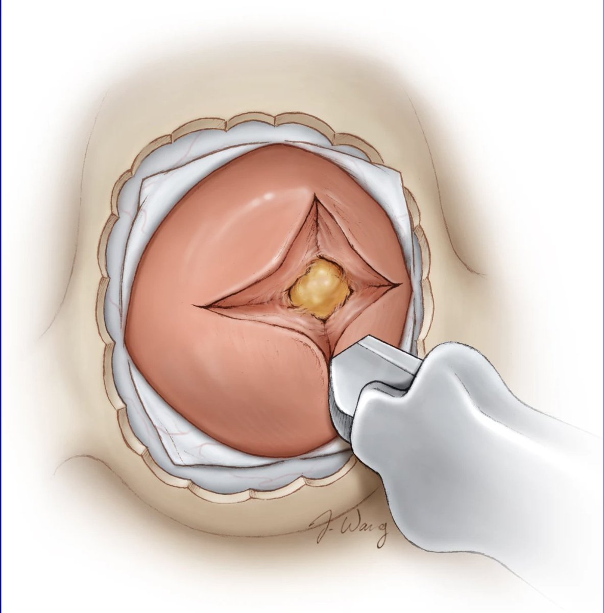
If the adenoma is not immediately seen when entering the gland, a Hardy dissector may be used to apply lateral pressure within the incision to force the microadenoma into the surgical field. Note the color difference between the microadenoma and the gland. Learn more here:

If the adenoma is not immediately seen when entering the gland, a Hardy dissector may be used to apply lateral pressure within the incision to force the microadenoma into the surgical field. Note the color difference between the microadenoma and the gland.

Learn more here:
Neurosurgical Atlas (@neurosurgatlas) 's Twitter Profile Photo

In this operative video, Dr. Cohen demonstrates the anterior petrosal approach for resection of a recurrent superior petroclival epidermoid cyst. After elevating the temporal lobe and identifying the incisura, careful dissection was performed around the fourth and fifth nerves

Neurosurgical Atlas (@neurosurgatlas) 's Twitter Profile Photo

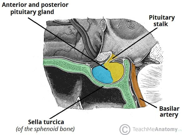
When operating on a microadenoma, incomplete removal of what key landmark separating the pituitary gland from the sphenoid sinus can lead to incomplete tumor resection? #Neurosurgery #EndoscopicSurgery #PituitaryTumor #Neuroanatomy

When operating on a microadenoma, incomplete removal of what key landmark separating the pituitary gland from the sphenoid sinus can lead to incomplete tumor resection? 

#Neurosurgery #EndoscopicSurgery #PituitaryTumor #Neuroanatomy
Neurosurgical Atlas (@neurosurgatlas) 's Twitter Profile Photo

The bone of the sella turcica plays a critical role in both accessing and successfully removing pituitary adenomas. Inadequate bone removal is the most common cause of subtotal tumor resection. Learn more here: zurl.co/04MJB #Neurosurgery #EndoscopicSurgery

The bone of the sella turcica plays a critical role in both accessing and successfully removing pituitary adenomas. Inadequate bone removal is the most common cause of subtotal tumor resection.

Learn more here: zurl.co/04MJB

#Neurosurgery #EndoscopicSurgery
Neurosurgical Atlas (@neurosurgatlas) 's Twitter Profile Photo

In this operative video, Dr. Cohen demonstrates the management of intraoperative rupture during microsurgical clipping of an anterior communicating artery aneurysm using the supraorbital approach. After careful dissection, the optic nerve, carotid artery, and both A1 segments

Neurosurgical Atlas (@neurosurgatlas) 's Twitter Profile Photo

In this operative video, Dr. Cohen demonstrates the endoscopic endonasal resection of a tuberculum sellae meningioma. After dural opening, the tumor was dissected off the right optic nerve. Further tumor decompression exposed the ACoA complex, and resection was further completed

Neurosurgical Atlas (@neurosurgatlas) 's Twitter Profile Photo

Why is autologous fat used to pack the resection cavity after pituitary tumor removal, and what complications can arise if the sella is overpacked? #Neurosurgery #EndoscopicSurgery #PituitaryTumor #Neuroanatomy

Why is autologous fat used to pack the resection cavity after pituitary tumor removal, and what complications can arise if the sella is overpacked? 

#Neurosurgery #EndoscopicSurgery #PituitaryTumor #Neuroanatomy
Neurosurgical Atlas (@neurosurgatlas) 's Twitter Profile Photo
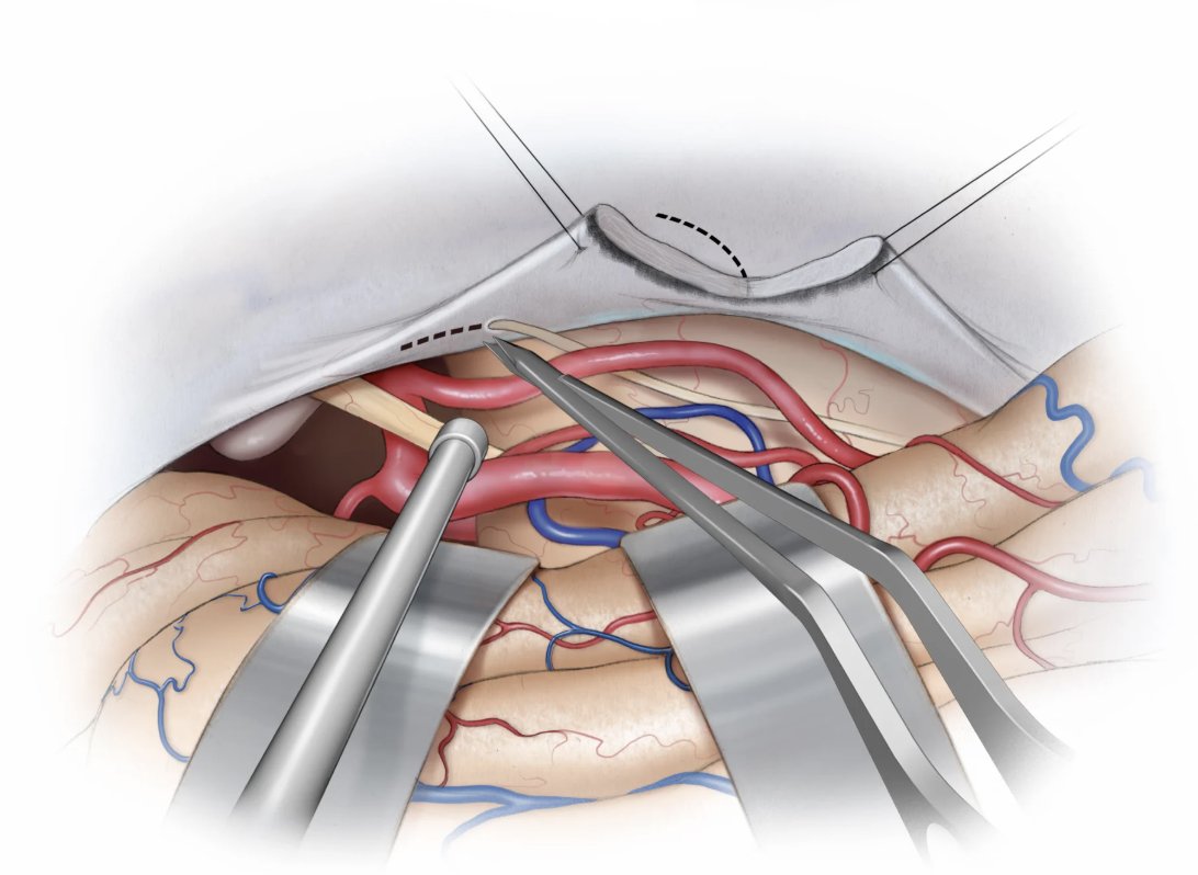
Autologous fat is used to pack the resection cavity after pituitary tumor removal to seal small cerebrospinal fluid (CSF) leaks caused by disruption of the diaphragma sellae and to support the remaining gland. Overpacking may cause chiasmal compression. Learn more

Autologous fat is used to pack the resection cavity after pituitary tumor removal to seal small cerebrospinal fluid (CSF) leaks caused by disruption of the diaphragma sellae and to support the remaining gland. Overpacking may cause chiasmal compression. 

Learn more
Dr. Z Neurosurgery (@drzneurosurgery) 's Twitter Profile Photo

A precise dissection of the temporal bone anatomy highlighting the chrodae tympani, jugular bulb, and segments of the internal carotid artery. Vital knowledge for safe and effective skull base and ear surgeries. #Anatomy #Neurosurgery #MedicalEducation

A precise dissection of the temporal bone anatomy highlighting the chrodae tympani, jugular bulb, and segments of the internal carotid artery. Vital knowledge for safe and effective skull base and ear surgeries.
#Anatomy #Neurosurgery #MedicalEducation