@patologiaymedicinabucal🔬👅 (@patoymedbucal) 's Twitter Profile
@patologiaymedicinabucal🔬👅

@patoymedbucal

UCVista, profesora de Histologia UCV, Patología y Medicina Bucal. Venezuela #OralPathology #PatologiaBucal #OralPath #OralMedicine #MedicinaBucal #MedBucal

ID: 2427814692

calendar_today04-04-2014 20:34:55

10,10K Tweet

6,6K Followers

4,4K Following

Head Neck Pathol (@headneckpathol) 's Twitter Profile Photo

“So You Made a Mistake” – The Path Forward Through Surgical Pathology Errors by Extreme Ownership and a Focus on the Patient James S. Lewis Jr. rdcu.be/ewSW3 #HeadNeckPathol #HNPJ #HeadandNeckPath #Pathology #ENTPath #PathologyTweet

“So You Made a Mistake” – The Path Forward Through Surgical Pathology Errors by Extreme Ownership and a Focus on the Patient
James S. Lewis Jr.
rdcu.be/ewSW3
#HeadNeckPathol #HNPJ #HeadandNeckPath #Pathology #ENTPath #PathologyTweet
María Laura García (@atusaludenlinea) 's Twitter Profile Photo

El Dr. Javier Soteldo, destacado oncólogo y científico venezolano, ha sido reconocido como el investigador número uno de Venezuela en el prestigioso AD Scientific Index 2025, un ranking internacional que evalúa a más de 2.5 millones de científicos en todo el mundo. Además, el Dr.

El Dr. Javier Soteldo, destacado oncólogo y científico venezolano, ha sido reconocido como el investigador número uno de Venezuela en el prestigioso AD Scientific Index 2025, un ranking internacional que evalúa a más de 2.5 millones de científicos en todo el mundo. Además, el Dr.
Tim Bracey (@drtimbracey) 's Twitter Profile Photo

Buccal mucosa white patch. This is severe dysplasia p16+ with transcriptionally active HR-HPV. In contrast to the counterpart in oropharynx implications of HPV+ in oral cavity neoplasia is less clear and management is not currently altered by this finding #ENTpath #PathTwitter

Buccal mucosa white patch. This is severe dysplasia p16+ with transcriptionally active HR-HPV. In contrast to  the counterpart in oropharynx implications of HPV+ in oral cavity neoplasia is less clear and management is not currently altered by this finding #ENTpath #PathTwitter
Lester Thompson (@headandneckpath) 's Twitter Profile Photo

Eosinophils and the Head and Neck Henok Eskinder & Yang Zhang rdcu.be/ewSXP #HeadNeckPathol #HNPJ #HeadandNeckPath #Pathology #ENTPath #PathologyTweet

Eosinophils and the Head and Neck
Henok Eskinder & Yang Zhang
rdcu.be/ewSXP
#HeadNeckPathol #HNPJ #HeadandNeckPath #Pathology #ENTPath #PathologyTweet
NotasDePatologia (@notas_patologia) 's Twitter Profile Photo

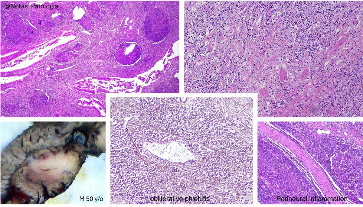
IgG4-Related Disease and Autoimmune Pancreatitis: Updated Review 1. Introduction IgG4-related disease (IgG4-RD) is a systemic immune-mediated fibroinflammatory condition characterized by infiltration of IgG4-positive plasma cells, storiform-type fibrosis, and obliterative

IgG4-Related Disease and Autoimmune Pancreatitis: Updated Review

1. Introduction

IgG4-related disease (IgG4-RD) is a systemic immune-mediated fibroinflammatory condition characterized by infiltration of IgG4-positive plasma cells, storiform-type fibrosis, and obliterative
NotasDePatologia (@notas_patologia) 's Twitter Profile Photo

🧠 Mantle Cell Lymphoma (MCL): Pathology Review with Emphasis on Lymphomatous Polyposis and Aggressive Variants 📌 Definition Mantle cell lymphoma (MCL) is a mature B-cell neoplasm derived from the follicular mantle zone. It is typically associated with the t(11;14)(q13;q32)

🧠 Mantle Cell Lymphoma (MCL): Pathology Review with Emphasis on Lymphomatous Polyposis and Aggressive Variants

📌 Definition

Mantle cell lymphoma (MCL) is a mature B-cell neoplasm derived from the follicular mantle zone. It is typically associated with the t(11;14)(q13;q32)
NotasDePatologia (@notas_patologia) 's Twitter Profile Photo

📌 Pulmonary Actinomycosis: The Great Mimicker of Lung Cancer 🔬 A Diagnostic Challenge in Respiratory Medicine Pulmonary actinomycosis is an uncommon, chronic granulomatous infection caused by anaerobic or microaerophilic Gram-positive bacilli of the genus Actinomyces,

📌 Pulmonary Actinomycosis: The Great Mimicker of Lung Cancer
🔬 A Diagnostic Challenge in Respiratory Medicine

Pulmonary actinomycosis is an uncommon, chronic granulomatous infection caused by anaerobic or microaerophilic Gram-positive bacilli of the genus Actinomyces,
@patologiaymedicinabucal🔬👅 (@patoymedbucal) 's Twitter Profile Photo

Esta es una de las cosas que más me gusta de la docencia, tienes la posibilidad de cambiar vidas y eso es al menos para mí algo maravilloso.

Janira Navarro (@janiranavarro) 's Twitter Profile Photo

Aberrant staining patterns for E-cadherin IHC -Cytoplasmic -Nuclear -Dot like perinuclear -Complete but week membranous staining -Fragmented, focal or beaded membranous Dr. Yantis-The Best of USCAP Journals #USCAP25 #pathology #PathX

Aberrant staining patterns for E-cadherin IHC
-Cytoplasmic
-Nuclear
-Dot like perinuclear
-Complete but week membranous staining
-Fragmented, focal or beaded membranous

Dr. Yantis-The Best of USCAP Journals
#USCAP25 #pathology #PathX
Ziad El-Zaatari (@ziad_zaatari) 's Twitter Profile Photo

🔬 Parotid Gland Tumor ~ Can you make the diagnosis at super low power? Or is it #TooLowToDiagnose ? #Pathology #ENTpath #PathResidents

🔬 Parotid Gland Tumor ~ Can you make the diagnosis at super low power? Or is it #TooLowToDiagnose ? #Pathology #ENTpath #PathResidents
NotasDePatologia (@notas_patologia) 's Twitter Profile Photo

Oncocytic Mucoepidermoid Carcinoma of the Parotid Gland: Comprehensive Review Definition Oncocytic mucoepidermoid carcinoma (OMEC) is an extremely rare variant of mucoepidermoid carcinoma (MEC), characterized by the predominance (>50%) of oncocytic cells, along with the

Oncocytic Mucoepidermoid Carcinoma of the Parotid Gland: Comprehensive Review

Definition

Oncocytic mucoepidermoid carcinoma (OMEC) is an extremely rare variant of mucoepidermoid carcinoma (MEC), characterized by the predominance (>50%) of oncocytic cells, along with the
NotasDePatologia (@notas_patologia) 's Twitter Profile Photo

Warthin-like Mucoepidermoid Carcinoma of the Salivary Gland Mucoepidermoid carcinoma (MEC) is the most common malignant salivary gland tumor. The Warthin-like variant (WL-MEC) is a rare morphological subtype recently recognized in the WHO Classification of Head and Neck Tumours

Warthin-like Mucoepidermoid Carcinoma of the Salivary Gland

Mucoepidermoid carcinoma (MEC) is the most common malignant salivary gland tumor. The Warthin-like variant (WL-MEC) is a rare morphological subtype recently recognized in the WHO Classification of Head and Neck Tumours
Lester Thompson (@headandneckpath) 's Twitter Profile Photo

Pitfalls in Diagnosis of Myoepithelial Carcinoma of Salivary Glands: A Study of 3 Cases with Cytologic-histologic Correlation and Molecular Analysis Shweta Agarwal rdcu.be/ewSYe #HeadNeckPathol #HNPJ #HeadandNeckPath #Pathology #ENTPath #PathologyTweet

Pitfalls in Diagnosis of Myoepithelial Carcinoma of Salivary Glands: A Study of 3 Cases with Cytologic-histologic Correlation and Molecular Analysis
Shweta Agarwal
rdcu.be/ewSYe
#HeadNeckPathol #HNPJ #HeadandNeckPath #Pathology #ENTPath #PathologyTweet
Springer Pathology (@springerpath) 's Twitter Profile Photo

Research in Head Neck Pathol reveals that Ki-67 labeling is significantly higher in oral squamous cell carcinoma compared to oral leukoplakia. The study also notes differences in 8-OHdG expression related to dysplasia severity in oral leukoplakia cases. bit.ly/40LyFqD

Springer Pathology (@springerpath) 's Twitter Profile Photo

Histopathological findings from a case of a 13-year-old male reveal a dedifferentiated solitary fibrous tumor in the lower jaw, characterized by spindle cells and specific immunohistochemical markers. The patient faced disease progression post-surgery. bit.ly/4mulSBf

Rossana Melo (@rossanamelo5) 's Twitter Profile Photo

Here's a high-resolution peek at #neutrophils isolated straight from the human bloodstream, captured in stunning detail with #electronmicroscopy and colorized! 🤩 #MicroscopyMonday

Here's a high-resolution peek at #neutrophils isolated straight from the human bloodstream, captured in stunning detail with #electronmicroscopy and colorized! 🤩 #MicroscopyMonday