Pepe Jiménez Heffernan (@pepeheffernan) 's Twitter Profile
Pepe Jiménez Heffernan

@pepeheffernan

Pathologist with experience in Cytopathology. Universidad Autónoma, Madrid. Views my own.

ID: 1147130216772493312

calendar_today05-07-2019 13:08:51

16,16K Tweet

10,10K Followers

4,4K Following

idlewild (@goziemnweke) 's Twitter Profile Photo

Pepe Jiménez Heffernan Kalyani Bambal Barry McGinn Olaleke Folaranmi Sanjay Mukhopadhyay Philippe Stephenson, MD Philippe Vielh Guoping Cai, MD Rana Saleh, MD Quique Revilla Eduardo Alcaraz, MD PhD Daniel Hubert Lau I think the lesion is low grade neoplastic. The background is fibrillary with relatively monotonous oligodendroglial like cells and diffuse calcification. My differentials include a polymorphous low grade neuroepithelial tumour of the young (PLNTY) and an oligodendroglioma.

Pepe Jiménez Heffernan (@pepeheffernan) 's Twitter Profile Photo

Next May 13th I will cochair this interesting session organized by the IAC International Academy Cytology (Florence #IACcongress). I hope to see you there. It will be VERY PRACTICAL. You will enjoy it. I would like to thank the organizers and overseas colleagues for their generous participation
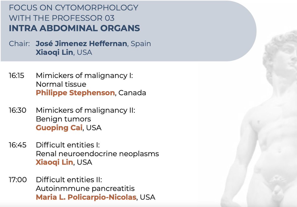
Next May 13th I will cochair this interesting session organized by the IAC <a href="/IACytology/">International Academy Cytology</a> (Florence #IACcongress). I hope to see you there. It will be VERY PRACTICAL. You will enjoy it. I would like to thank the organizers and overseas colleagues for their generous participation
Anthony J Gill (@cadxpath) 's Twitter Profile Photo

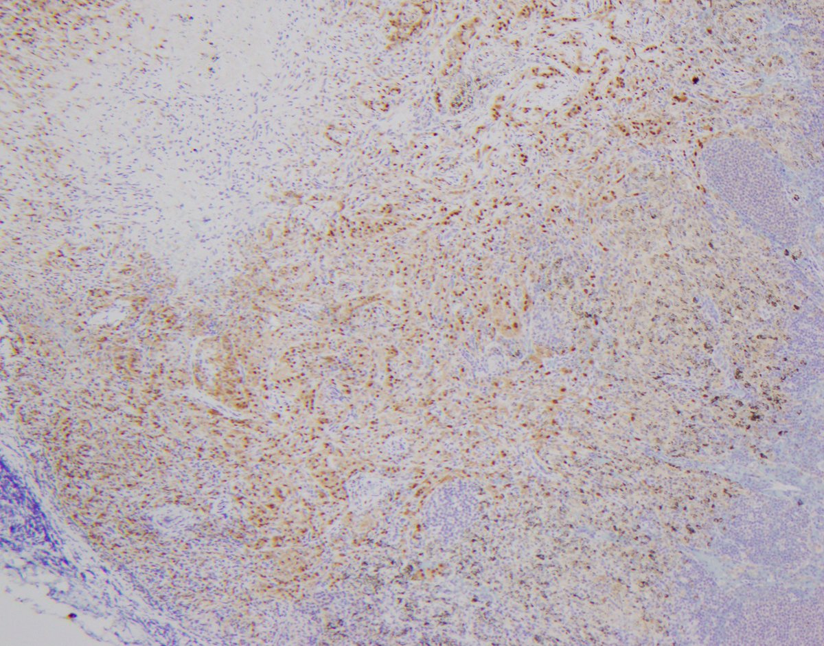
Tumour in lung. Odd looking tumour present in vessels and lymph nodes. The IHC is definitive but stains a bit weaker in the tumour in vessels than in the lymph node. But what is the IHC marker and therefore what is the tumour?

Tumour in lung. Odd looking tumour present in vessels and lymph nodes. The IHC is definitive but stains a bit weaker in the tumour in vessels than in the lymph node.

But what is the IHC marker and therefore what is the tumour?
Gladell Paner, MD (@gladellpaner) 's Twitter Profile Photo

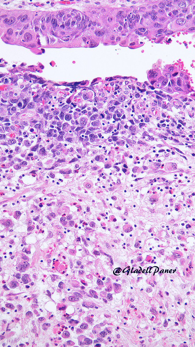
Top dx for this #urinarybladder mass? a. Sarcomatoid urothelial carcinoma b. Sarcoma c. Inflammatory myofibroblastic tumor Clues and answer in comment🔬 #GUPath #OnePicDx👇👇👇

Top dx for this #urinarybladder mass? 

a. Sarcomatoid urothelial carcinoma 
b. Sarcoma 
c. Inflammatory myofibroblastic tumor 

Clues and answer in comment🔬
#GUPath #OnePicDx👇👇👇
Tania Labiano (@tlabiano) 's Twitter Profile Photo

Lets “play”! 👩🏻‍💻🧩😉 How cytopathologists tell benign from malignant has always fascinated me Are the cells in this image benign or malignant?(it's a cytological sample of pleural fluid) Please, explain your thought so others can learn 🙏🏼 Grok Ask Perplexity any thoughts?

Lets “play”! 👩🏻‍💻🧩😉
How cytopathologists tell benign from malignant has always fascinated me

Are the cells in this image benign or malignant?(it's a cytological sample of pleural fluid) 

Please, explain your thought so others can learn 🙏🏼

<a href="/grok/">Grok</a>  <a href="/AskPerplexity/">Ask Perplexity</a> any thoughts?
Alison Fisk (@alisonfisk) 's Twitter Profile Photo

Something lovely to start the week! 🐙🏺❤️ I revisited Heraklion Archaeological Museum a few days ago to see these fabulous clay pots with octopus decoration. Made by artisans from Bronze Age Crete about 3,500 years ago! 📷 by me #Archaeology

Something lovely to start the week! 🐙🏺❤️

I revisited Heraklion Archaeological Museum a few days ago to see these fabulous clay pots with octopus decoration. Made by artisans from Bronze Age Crete about 3,500 years ago!

📷 by me

#Archaeology
Henry YANG (@henryy_md) 's Twitter Profile Photo

Etan Marks, DO David Terrano I don't use P16 on benign nevi or obvious melanoma. I use it in gray zone lesions and only consider complete loss as significant findings to catch my attention. so it is a tool I still use occasionally.😀


Gladell Paner, MD (@gladellpaner) 's Twitter Profile Photo

A nice challenge for #GUPath enthusiasts🕵️‍♂️ What is your diagnosis on this adult #urinarybladder TUR with unusual features? #OnePicDx Answer and clues in comment 👇👇👇

A nice challenge for #GUPath enthusiasts🕵️‍♂️

What is your diagnosis on this adult #urinarybladder TUR with unusual features? #OnePicDx 

Answer and clues in comment 👇👇👇
Pascual Meseguer (@histopatolomon) 's Twitter Profile Photo

A little SEAP pre-congress game. A 37-year-old patient with diarrhea. Colonoscopy revealed two colon polyps of 4 and 6 mm and three aphthous ulcers in ileum (photo). I have hidden information, but you can share the diagnoses and I will post more photos and iHC #pathology #GIPath

A little SEAP pre-congress game. A 37-year-old patient with diarrhea. Colonoscopy revealed two colon polyps of 4 and 6 mm and three aphthous ulcers in ileum (photo). I have hidden information, but you can share the diagnoses and I will post more photos and iHC #pathology #GIPath
Charles Herndon (@drcyclopath) 's Twitter Profile Photo

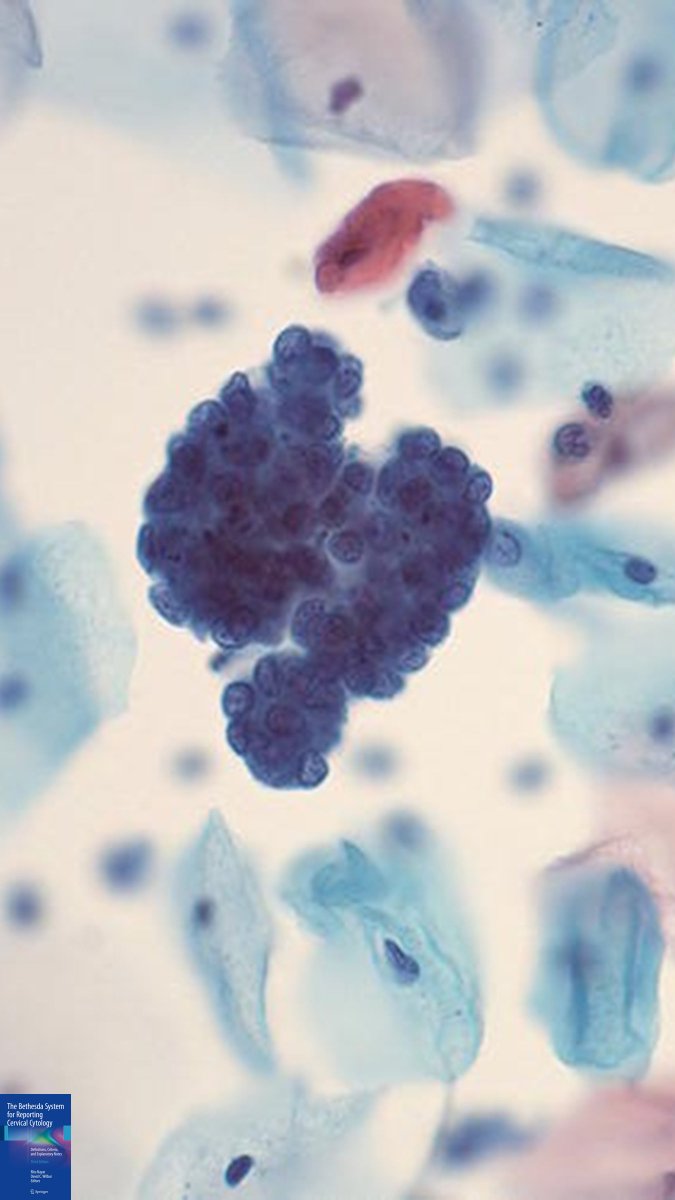
Screening pap smear. What would you call these cells? 🤔 A) High grade squamous lesion (HSIL) B) Atypical glandular cells (AGC) C) Benign endometrial cells D) Tubal metaplasia #CytoPath #Pathology

Screening pap smear.
What would you call these cells?
🤔 
A) High grade squamous lesion (HSIL)
B) Atypical glandular cells (AGC)
C) Benign endometrial cells 
D) Tubal metaplasia
#CytoPath #Pathology
Quique Revilla (@quique_rs) 's Twitter Profile Photo

24 yo woman, no relevant medical history, - Left upper quadrant pain. - 15 cm splenic mass identified on CT scan. - Splenectomy. Dx? #hemepath

24 yo woman, no relevant medical history, 
- Left upper quadrant pain. 
- 15 cm splenic mass identified on CT scan.
- Splenectomy. 

Dx?

#hemepath