Seham Ebrahim (@sehamebrahim) 's Twitter Profile
Seham Ebrahim

@sehamebrahim

Assistant Professor @UVA. Interested in molecular mechanosensors. High-resolution microscopy: light, live and cryo-EM.

ID: 35974383

linkhttp://www.med.virginia.edu/ebrahim-lab calendar_today28-04-2009 03:28:23

123 Tweet

229 Followers

259 Following
Natalie Porat-Shliom (@poratshliomlab) 's Twitter Profile Photo

Preprint alert!! How zonated hepatocytes adapt to changes in lipid influx due to fasting or a Western diet (WD). We developed a microscopy-based phenotyping method called single-cell phenomics (scPhenomics) to measure the features of mitochondria and lipid droplets (LDs).

Patrick Oakes (@pwoakes) 's Twitter Profile Photo

I'm spending more time on the other social network these days, but it's still worth posting that we'll be at #cellbio24 #ASCB! If you like LIM proteins or septins come check out our posters:

I'm spending more time on the other social network these days, but it's still worth posting that we'll be at  #cellbio24 #ASCB! If you like LIM proteins or septins come check out our posters:
Sean R. Moore, MD (@moore_cchmc_gi) 's Twitter Profile Photo

Preprint alert: Ready-to-use supplemental foods save lives. Fascinating work by Zehra Jamil Aga Khan University Gabe Hanson Virginia BME and team reveals host-microbiome factors that predict degree + durability of response in childhood wasting Asad Ali MBBS, MPH AKU Community Health Sciences medrxiv.org/content/10.110…
Alushin_Lab (@alushin_lab) 's Twitter Profile Photo

Excited that the peer-reviewed version of our fascin structure paper is now out in NatureStructMolBiol. Thanks very much to the referees for their constructive feedback. rdcu.be/d6Tct

Dominik Robak (@dominikprobak) 's Twitter Profile Photo

Happy #MicroscopyMonday! With Seham Ebrahim we’re capturing single-cell behavior deep in the mouse intestine—no lumen opening needed! Thanks to neutrophil-YFP mice from Dr. Arandjelovic at UVA, we imaged “tiny steps” of neutrophils across endothelial cells at just 30 ms exposure

Seham Ebrahim (@sehamebrahim) 's Twitter Profile Photo

Have cutting-edge cell biology research to share? Submit to Molecular Biology of the Cell (MBoC)- dedicated to advancing discovery and fostering collaboration. Proud to be part of the editorial team supporting groundbreaking science. molbiolcell.org #CellBiology #MBoC

Dominik Robak (@dominikprobak) 's Twitter Profile Photo

#3DThursday with Seham Ebrahim Inside the gut: Villi = nutrient uptake + barrier + immune balance. 🔴 Phalloidin 🔵 DAPI 🟢 Clvd caspase-3 (apoptotic extrusion!) Fixed tissue, imaged in 3D — stunning, structural, and full of insight. #Microscopy #3DImaging #Biophysics

Natalie Porat-Shliom (@poratshliomlab) 's Twitter Profile Photo

Have you ever wondered if liver zonation is conserved from mice to humans? And how does fibrosis affect zonation in patients? Now, these questions are answered using scDVP. Some collaborations are meant to be Caroline Weiss Florian Rosenberger Matthias Mann Lab MLSB

Manuela Casasoli (@manuelacasasoli) 's Twitter Profile Photo

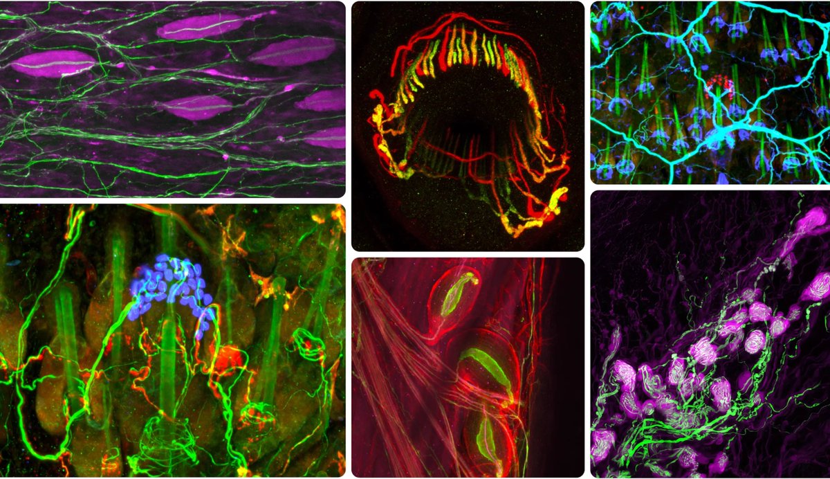
The neurons beneath everyday sensations. "How does pressure and vibration across millions of nerve endings become a hug?" "Of all the senses, the somatosensory system is the most complex, and therefore touch, some researchers argue, is the most difficult to study." #Science

The neurons beneath everyday sensations.

"How does pressure and vibration across millions of nerve endings become a hug?"

"Of all the senses, the somatosensory system is the most complex, and therefore touch, some researchers argue, is the most difficult to study."

#Science
Dominik Robak (@dominikprobak) 's Twitter Profile Photo

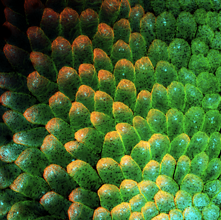
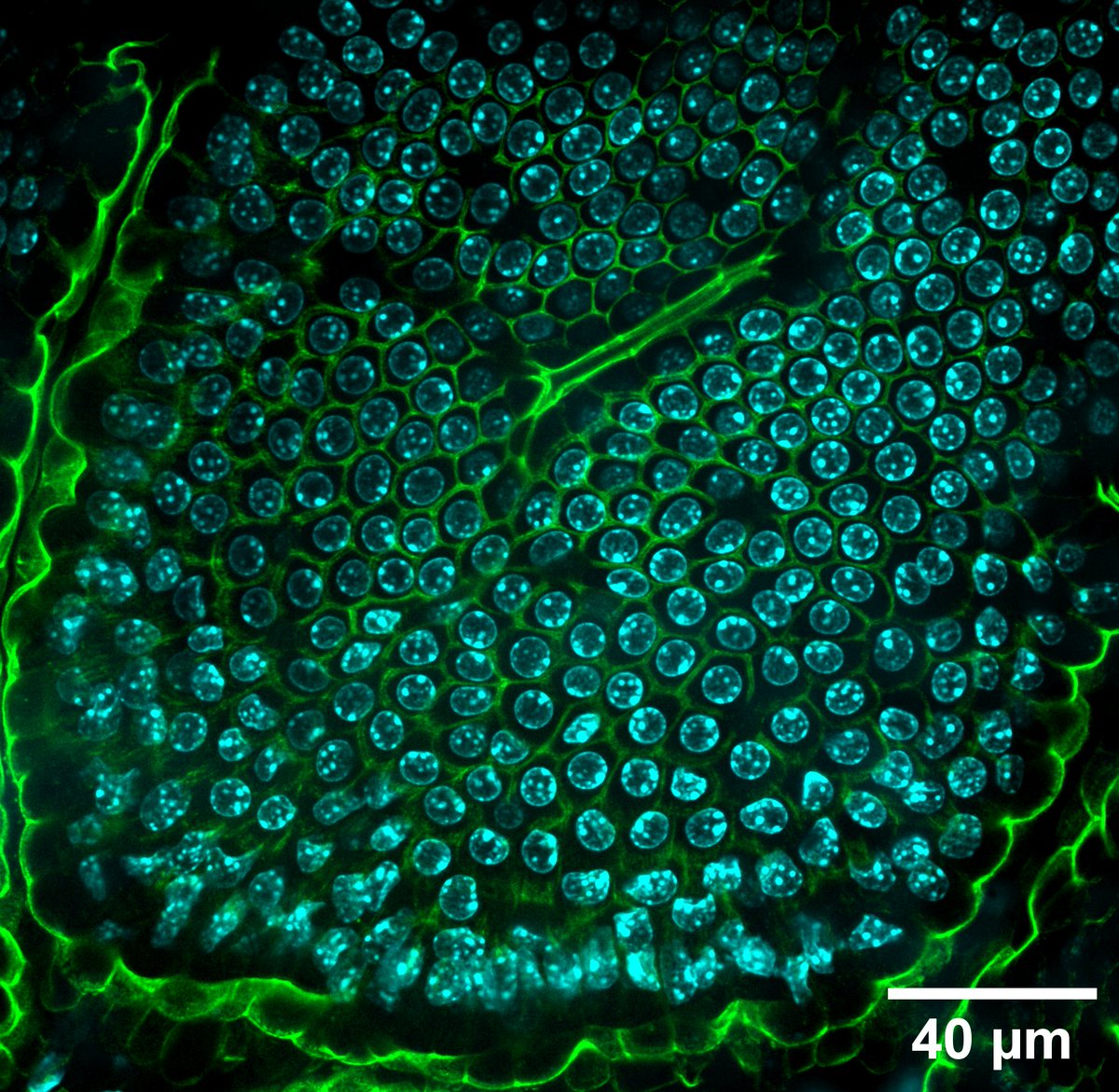
Happy #MicroscopyMonday! Bees didn’t invent the honeycomb—biology’s been doing it at the single-cell level all along. 🟢 GFP-tagged junctions 🔵 Hoechst-stained nuclei A living mouse intestinal villus. No blueprint. Just (bio)physics. #intravitalimaging #cellbiology #OME

Happy #MicroscopyMonday!

Bees didn’t invent the honeycomb—biology’s been doing it at the single-cell level all along.

🟢 GFP-tagged junctions
🔵 Hoechst-stained nuclei

A living mouse intestinal villus.

No blueprint. Just (bio)physics.

#intravitalimaging #cellbiology #OME
Seham Ebrahim (@sehamebrahim) 's Twitter Profile Photo

From stem cell to structure- This human colonoid shows the self-organization of intestinal epithelial cells in 3D—nuclei in magenta, actin in green. A living miniature gut for your #FluorescenceFriday.

From stem cell to structure-
This human colonoid shows the self-organization of intestinal epithelial cells in 3D—nuclei in magenta, actin in green. A living miniature gut for your #FluorescenceFriday.
John Damianos, M.D. (@john_damianosmd) 's Twitter Profile Photo

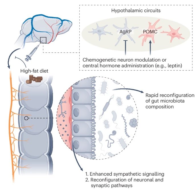
We know the #microbiome can influence the brain, but the brain can also influence the microbiome! #MedTwitter #GITwitter nature.com/articles/s4225…

We know the #microbiome can influence the brain, but the brain can also influence the microbiome! 

#MedTwitter #GITwitter 

nature.com/articles/s4225…
Seham Ebrahim (@sehamebrahim) 's Twitter Profile Photo

"Stereocilia"- actin-based skyscrapers that adorn sensory cells of our inner ear and covert vibrations into electrical signals—the first step in hearing and balance. #FluorescenceFriday

"Stereocilia"- actin-based skyscrapers that adorn sensory cells of our inner ear and covert vibrations into electrical signals—the first step in hearing and balance. #FluorescenceFriday
Bechara Kachar (@becharakachar) 's Twitter Profile Photo

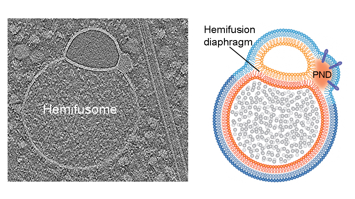
New organelle uncovered by in situ #cryoET: the #hemifusome, a stable, hemifused vesicle pair separated by an extended hemifusion diaphragm. This structure redefines what membrane fusion intermediates can do. rdcu.be/emtQu #CryoET #MembraneBiogenesis #Organelle 🧵1/5

New organelle uncovered by in situ #cryoET: the #hemifusome, a stable, hemifused vesicle pair separated by an extended hemifusion diaphragm. This structure redefines what membrane fusion intermediates can do.

rdcu.be/emtQu

#CryoET #MembraneBiogenesis #Organelle

🧵1/5
Amirrasoul Tavakoli (@amirrasoultav) 's Twitter Profile Photo

Grateful to see a briefing of our new paper shared more widely always meaningful when the science resonates beyond the lab. Great collaboration with amazing scientists shiqiong hu Seham Ebrahim Bechara Kachar