JuanMiranda (@themskarchive) 's Twitter Profile
JuanMiranda

@themskarchive

Musculoskeletal Radiologist. Husband. Obsessive reader. Investor. Python. Grateful to share my daily MSK and Tanzanian activities #radiology #radtwitter #MSKRad

ID: 1599757835289632773

calendar_today05-12-2022 13:29:49

1,1K Tweet

2,2K Followers

114 Following

JuanMiranda (@themskarchive) 's Twitter Profile Photo

1/12/2023. CAPNON is one of the rarest pseudotumors of the neuroaxis. Here is a very interesting case manifested as an intrathecal lesion in the lumbar canal. Post-qx and histopathology details included. Full details here: ajnr.org/content/com/20…

1/12/2023. CAPNON is one of the rarest pseudotumors of the neuroaxis. Here is a very interesting case manifested as an intrathecal lesion in the lumbar canal. Post-qx and histopathology details included. Full details here: ajnr.org/content/com/20…
JuanMiranda (@themskarchive) 's Twitter Profile Photo

25/1/2024. 1⃣What is the likely mechanism of this injury and the name? 2⃣What is the name of the ligament affected, and 3⃣what other intra-articular lesions would you expect to find in this patient's #knee #MRI? 🦵🦵

25/1/2024. 1⃣What is the likely mechanism of this injury and the name? 2⃣What is the name of the ligament affected, and 3⃣what other intra-articular lesions would you expect to find in this patient's #knee #MRI? 🦵🦵
JuanMiranda (@themskarchive) 's Twitter Profile Photo

5/4/2024. 🟠 23yo male presents to ED with left #knee 🦵contusion. #XRAY reveals: 1⃣ Superolateral patellar fracture, two displaced fragments. 2⃣ Type III tripartite patella. Let's dive into the diagnosis. MRI and rest of the findings incoming. #MSKrad #Trauma #OrthoTwitter

5/4/2024. 🟠  23yo male presents to ED with left #knee 🦵contusion. #XRAY reveals: 
1⃣ Superolateral patellar fracture, two displaced fragments.
2⃣ Type III tripartite patella.
Let's dive into the diagnosis. MRI and rest of the findings incoming.
#MSKrad #Trauma #OrthoTwitter
JuanMiranda (@themskarchive) 's Twitter Profile Photo

1/7/2024. 💪 A rare, but possible cause of axillary nerve entrapment 🟥: inferior ganglion cyst / paralabral glenohumeral cyst contacting with the axillary nerve in the cuadrangular space. No physical or MR signs of denervation. #MRI #shoulder #nerveentrapment #axillary

1/7/2024. 💪 A rare, but possible cause of axillary nerve entrapment 🟥: inferior ganglion cyst / paralabral glenohumeral cyst contacting with the axillary nerve in the cuadrangular space. No physical or MR signs of denervation.  #MRI #shoulder #nerveentrapment #axillary
JuanMiranda (@themskarchive) 's Twitter Profile Photo

9/1/2025. Bipartite patella Type I 🦴: a congenital variant with an unfused inferior patellar pole. On 🩻, look for a distinct fragment with smooth margins; MRI reveals a fibrocartilaginous bridge with mild degenerative changes. #Radiology #Ortho #BoneHealth

9/1/2025. Bipartite patella Type I 🦴: a congenital variant with an unfused inferior patellar pole. On 🩻, look for a distinct fragment with smooth margins; MRI reveals a fibrocartilaginous bridge with mild degenerative changes. #Radiology #Ortho #BoneHealth
JuanMiranda (@themskarchive) 's Twitter Profile Photo

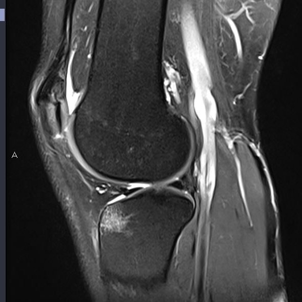
17/2/2024. 🦵This 20-year-old female presents with probable suprapatellar plica syndrome (thickened & non-fenestrated) + suprapatelar distended bursitis & synovial proliferation. 🟠#KneePain #Orthopedics #Radiology #MedicalImaging #SportsMedicine #PlicaSyndrome #Bursitis

17/2/2024. 🦵This 20-year-old female presents with probable suprapatellar plica syndrome (thickened & non-fenestrated) + suprapatelar distended bursitis & synovial proliferation. 🟠#KneePain #Orthopedics #Radiology #MedicalImaging #SportsMedicine #PlicaSyndrome #Bursitis
JuanMiranda (@themskarchive) 's Twitter Profile Photo

🔎DIAGNOSIS: 💀 X-ray: Calcifications in arcs and rings in the proximal humeral metaphysis, suggesting a cartilaginous matrix. 🧲 MRI: Popcorn-like lesion with characteristic signal and morphology of a low-grade chondral tumor, with no signs of aggressiveness. #Radiology #MSK

🔎DIAGNOSIS: 
💀 X-ray: Calcifications in arcs and rings in the proximal humeral metaphysis, suggesting a cartilaginous matrix.
🧲 MRI: Popcorn-like lesion with characteristic signal and morphology of a low-grade chondral tumor, with no signs of aggressiveness.

#Radiology #MSK
JuanMiranda (@themskarchive) 's Twitter Profile Photo

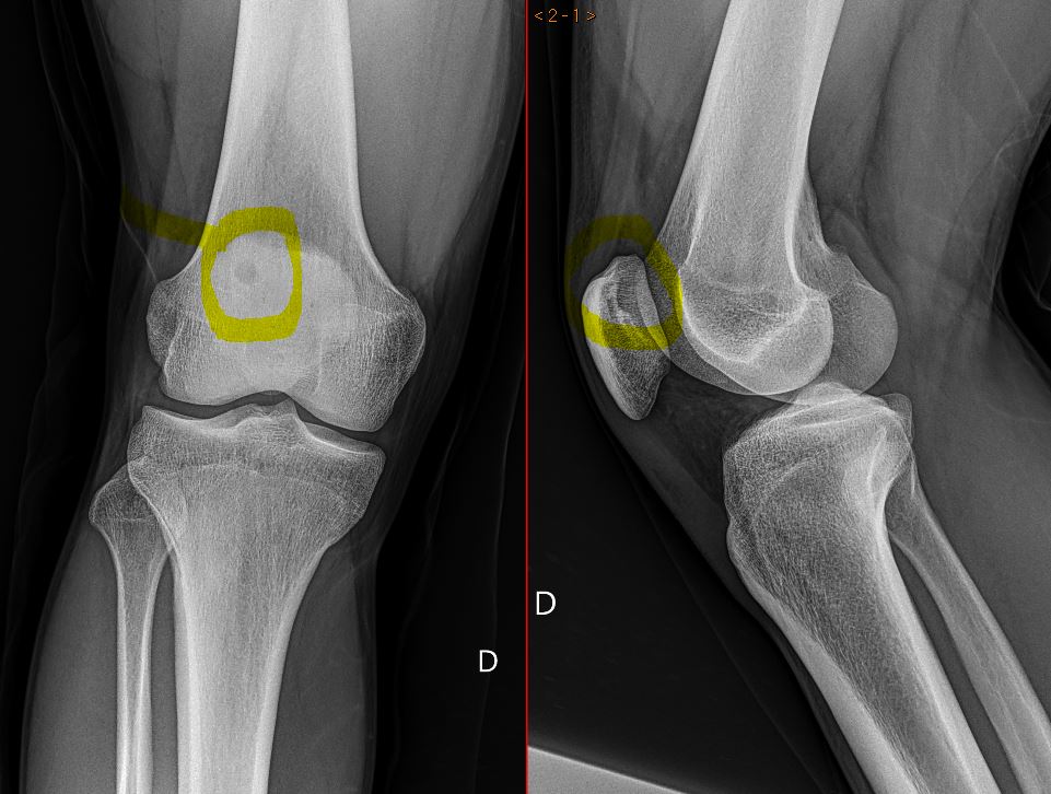
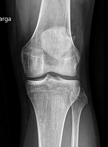
18/3/2025. ✅ Two views show a femoral lesion. What’s the likely diagnosis? “No-touch” lesion? Share your thoughts! #Radiology #Ortho #MedTwitter #BoneLesion #Diagnosis 🔎🦴

18/3/2025. ✅ Two views show a femoral lesion. What’s the likely diagnosis? “No-touch” lesion? Share your thoughts!

#Radiology #Ortho #MedTwitter #BoneLesion #Diagnosis
🔎🦴
JuanMiranda (@themskarchive) 's Twitter Profile Photo

20/3/2025.🟥What´s the name of this quite rare variant of anatomy in the knee? Likely the cause of pain of this 51 year-old patient. 👍 #MSK #Radiology #Orthopedics #MedicalTwitter #MRI

20/3/2025.🟥What´s the name of this quite rare variant of anatomy in the knee? Likely the cause of pain of this 51 year-old patient. 👍

#MSK #Radiology #Orthopedics #MedicalTwitter #MRI
JuanMiranda (@themskarchive) 's Twitter Profile Photo

Join us at the CIRME Ibero-American MSK Radiology Course – 4th Edition 🇪🇸 📍 Santander, May 16–17, 2025 💥 Imaging of sports injuries, guided interventions & more! 🔗 info.tecnoimagen.com.ar/cirme-2025-san… #MSKradiology #CIRME2025 #MedicalImaging #SportsMedicine #Ultrasound

Join us at the CIRME Ibero-American MSK Radiology Course – 4th Edition 🇪🇸

📍 Santander, May 16–17, 2025
💥 Imaging of sports injuries, guided interventions & more!
🔗 info.tecnoimagen.com.ar/cirme-2025-san…

#MSKradiology #CIRME2025 #MedicalImaging #SportsMedicine #Ultrasound